Effective procedures for the treatment of hyperkeratosis
Phototherapy
Plasma therapy
NeoGen procedure
Mesotherapy
Peeling
Medical pedicure
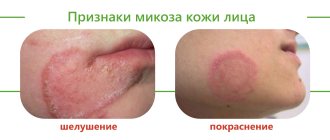
One of the fairly common types of skin diseases is hyperkeratosis. This is an excessive thickening of the stratum corneum of the epidermis. With the development of such a pathology, horn cells begin to rapidly divide and desquamation is impaired, which ultimately leads to thickening of the skin. Moreover, the thickening can reach several centimeters. According to statistics, hyperkeratosis is observed in approximately 20% of men and 40% of women. As a rule, the problem occurs in individual areas and leads to a significant deterioration in the appearance of the skin.
In children, the pathology can be observed at the age of 1–3 years, and it mainly affects the face and hands. In adults, changes in the stratum corneum are more common on the feet and toenails, as well as on the face and scalp. There are several types of the disease, which have their own symptoms and causes of development. Let's figure out what hyperkeratosis is and whether such a pathology is dangerous for health.
Classification
The classification of hyperkeratoses is based on a number of factors, according to which the following are distinguished:
- By origin: hereditary and acquired hyperkeratosis .
- By area of damage: local (calluses/warts) and diffuse, occupying large areas of the skin.
According to clinical forms:
- Follicular hyperkeratosis - in which exfoliating scales of the stratum corneum clog the ducts of the follicles, which is manifested by the appearance of numerous small tubercles on the skin.
- Seborrheic hyperkeratosis is localized mainly on the scalp, less often on the facial skin. It manifests itself in the formation of areas of peeling in the form of an easily detachable greasy crust, after which reddish spots remain.
- Warty hyperkeratosis . It is characterized by the appearance on the skin of formations that outwardly resemble warts, but without the etiological factor of true warts - papillomavirus .
- Hyperkeratosis of the feet . It manifests itself as thickening of the skin of the feet with the appearance of corns and calluses.
- Disseminated hyperkeratosis . It appears as polymorphic elements on the skin of the trunk and limbs, reminiscent of thick/short hair, located separately and not merging with each other.
- Lenticular hyperkeratosis . It is characterized by the appearance of horny papules on the hair follicles of the lower extremities, and after removal, small indentations remain on the skin.
- Senile hyperkeratosis . The appearance of dark keratinized spots on the skin of older people is typical.
Features of the pathology
The concept of hyperkeratosis comes from two Greek words: hyper – “many” and keratosis – “formation of keratin”. In fact, hyperkeratosis is not an independent disease - most often it is just a symptom of other diseases. For example, such a problem occurs with lichen, ichthyosis or erythroderma. However, thickening of the stratum corneum can sometimes appear in a healthy person: for example, the feet, elbows and knees are often susceptible to this problem.
Hyperkeratosis is considered a potentially life-threatening disease, but treatment is still recommended. Pathology can cause serious cosmetic defects, which often lead to social maladaptation and decreased self-esteem.
The basis of the stratum corneum of the skin is made up of horny plates, which contain the protein keratin. Typically, the layer includes 15–20 layers of horny scales; when the epidermis thickens, their number can reach 100.
Essentially, the stratum corneum is the end result of the division of keratinocytes of the epidermis, which is why it has a specific structure. It is based on horn cells held together by intercellular lipids. Normally, this layer of the epidermis is constantly renewed: the horny scales are peeled off and replaced with new cells.
The desquamation process starts in the basal layer of the skin, where keratinocytes are produced. After this, the cells gradually move to the upper layers, losing their nucleus and organelles and turning into flat scales, which develop into the stratum corneum.
Normally, the epidermal renewal cycle lasts about 21–28 days; with age it slows down and can reach 45–72 days. In addition, the state of cellular metabolism is influenced by various factors such as lifestyle, nutrition, and hormonal levels of the body.
With hyperkeratosis, the patient's production of proteolytic enzymes, which regulate the processes of destruction of protein bonds between the cells of the stratum corneum, is disrupted. As a result, there is no desquamation of old cells, and due to the accumulation of keratinocytes, a significant thickening of the epidermis occurs.
List of sources
- Bauman L. Cosmetic dermatology. Principles and practice. Per. from English Potekaeva N. N. M.: MEDpress-inform. 2012. 688 p.
- Afanasyev E.N. Mechanically induced hyperkeratoses of the foot // Plastic surgery and cosmetology. 2012. No. 4. pp. 644–661.
- Kholodilova N.A., Monakhov K.N. The use of basic care products in patients with impaired skin barrier // Russian Journal of Skin and Venereal Diseases. 2009. No. 6. pp. 68–69.
- Lomakina E. A. The role of the barrier function of the skin in the pathogenesis of some dermatoses // Modern problems of dermatovenereology, immunology and medical cosmetology. 2009, no. 2. pp. 87–90.
- Rodionov A. N. Dry skin. Dermatocosmetology. Lesions of the facial skin and mucous membranes. Diagnosis, treatment and prevention. SPb: Science and Technology. 2011. 911 p. pp. 63–69.
Types of pathology
The following forms of hyperkeratosis are distinguished:
- Follicular. This type of pathology is characterized by damage to the hair follicles. Most often, follicular hyperkeratosis appears in areas with dry, dehydrated skin. The reasons are heredity, vitamin deficiency, inflammatory skin diseases or neglect of personal hygiene. There are two forms of pathology: with follicular hyperkeratosis of type I, spiky nodules and plaques appear around the hair follicle, with type II - a hemorrhagic rash caused by a lack of vitamin C and K. Very often, the ducts of the hair follicles are clogged with blood or red pigment.
- Lenticular. This form is characterized by the appearance of small “plugs”, most often on the hair follicles, which look like keratinized spots. The reasons for the development are not exactly known, but most often the problem occurs in elderly patients. As a rule, the pathology is chronic, but without a tendency to regression.
- Disseminated. With this form, polymorphic elements appear on the skin, which outwardly resemble short and thick hair. They are isolated from each other and have no tendency to grow together.
- Seborrheic. This type of pathology is typical for oily skin and appears as a result of metabolic disorders. At an early stage, characteristic yellow-brown spots of a round shape appear, which gradually fade and turn into plaques. The lesions can be quite large in diameter.
- Warty. Externally, the warty form resembles warts, but their appearance is not caused by papillomavirus.
- Plantar. Hyperkeratosis of the feet can occur on the entire sole or only on the heel area. As a rule, this form is characterized by severe dryness and roughness of the skin, as well as noticeable thickening in the affected areas. There is a dry callus (a local lesion with clear boundaries of thickening of the stratum corneum), a core callus (a sharply limited area of thickening of a round shape of small size) and a soft callus (appears between the fingers as a result of increased humidity).
- Subungual. Quite often, subungual hyperkeratosis occurs, resulting from traumatic onychia or onychomycosis. It is characterized by rapid growth from the distal edge, which causes an enlargement of the nail plate.
Plantar hyperkeratoses: clinical picture, diagnosis, treatment
Leonardo da Vinci has the following statement: “The human foot is a work of art, consisting of 26 bones, 107 ligaments and 19 muscles.” The great artist, writer, scientist and thinker of the High Renaissance was certainly right in treating the foot as a work of art. The interaction of the bone and ligamentous apparatus with the muscular system is brought here to impeccable perfection. And one can only marvel at how, despite the colossal load that our feet experience every day, they combine functional reliability and aesthetic appeal. This is due not only to the muscles, ligaments and bones, but also to the skin, the structure of which on the feet is significantly different due to anatomical and physiological characteristics.
The human foot is a rather complex mechanism designed to hold the entire body in an upright position when standing and walking. This small element of the entire musculoskeletal system in terms of mass and size constantly has to withstand significant static and dynamic loads throughout a person’s life. These features determined the structure of the foot - the lowest part of the limbs. The part of the foot in direct contact with the ground is called the foot or sole, the upper side opposite to it is called the dorsum of the foot. The foot as a whole has an arched structure, thanks to its articulations it has mobility, flexibility and elasticity. Externally, the foot is divided into fore, middle and rear sections. The anterior section includes the toes and from the sole side the ball of the foot, the middle section the arch of the foot, and the rear section from the sole side forms the heel. The arch is that part of the foot that normally does not touch the ground on the side of the sole, but on the back side forms the instep of the foot. According to the bone structure, the foot is divided into tarsus, metatarsus and phalanges. The convex part of the arch is made up of five metatarsal bones located in the body of the foot; the outer extensions of these bones form the fingers and are called phalanges. The ball of the foot is located at the lowest part of the arch in front of the toes and protects the joints from impact [1].
The heel bone is the strongest and heaviest of all 26 bones in the foot. It is she who is a continuation of the axis of the human body, and therefore it bears all his weight. Just like the heel, 6 more bones of the foot (tarsal bones) have a spongy structure, that is, inside they are almost completely filled with strong bone tissue, allowing them to withstand heavy loads. The remaining bones of the foot look like light hollow tubes of varying lengths. Their main task is to ensure mobility and shock-absorbing properties of the foot when walking, jumping and running. All bones of the foot have articular surfaces covered with smooth and slippery cartilage tissue, facilitating their mutual friction. The joints of the hind and midfoot are less mobile than the joints of the toes. Each joint is covered with a capsule, inside which a small amount of fluid is constantly formed, which promotes additional sliding of the articular surfaces of the bones.
Etiology and pathogenesis of plantar hyperkeratoses
The skin of the sole is thick, rough, hairless and rich in sweat glands. The skin on the dorsal surface is elastic and easily shifts, so with any inflammatory processes, swelling appears on the dorsum of the foot. A distinctive feature of the skin of the soles is that in this area there is the thickest epidermis, which, like on the palms, consists of five layers: basal, spinous, granular, shiny and horny. It is worth noting that the stratum lucidum is found only in the epidermis of the palms and soles. The keratinocytes of this layer contain a specific protein, eleidin, an intermediate product of the transformation of keratohyalin into keratin, which gives a characteristic shine upon histological examination. In places that serve to support bones: on the heel, on the heads of the metatarsal bones, on the nail phalanges, between the bones and the outer integument, there is a fairly well-defined third layer of skin - subcutaneous adipose tissue, which protects the bone from external pressure. At the level of the metatarsal heads, the transverse margin is a fat pad, also called the ball of the foot. A deep fold traces it in front of the plantar surface of the fingers, interrupted by individual interdigital spaces. This makes the fingers appear shorter on the side of the sole in relation to their size on the back side.
Under constant stress, with foot deformities, wearing uncomfortable shoes, and active sports, in response to mechanical stress on the skin of the feet, a response occurs in the form of increased proliferation of keratinocyte cells, which ultimately leads to the development of plantar hyperkeratosis. The concept of “hyperkeratosis” comes from two Greek words: ὑπέρ - many and keratosis - formation of keratin. Hyperkeratosis makes the skin rigid, less elastic, and reduces its sensitivity to external influences. There are many reasons that cause plantar hyperkeratosis. The main ones are presented in Table 1.
Hyperkeratoses caused by mechanical causes are among the most common among healthy young people involved in sports, as well as among the elderly and patients with chronic diseases. According to the literature, more than half of people over 65 years of age and more than 65% of patients with rheumatoid arthritis have hyperkeratoses that require treatment [1]. According to Springett, when examining men and women of all age groups at an outpatient appointment, hyperkeratosis is detected in the area of the 1st metatarsophalangeal joint (MTP) in 27% of cases, 2–4 MTP joints in 36% and 5 MTP joints in 17% of cases. In the Grouios studies, men involved in running took part, and approximately comparable results were obtained: hyperkeratosis of the 1st PFJ was detected in 23%, 2–4 PFJs in 32%, and 5 PFJs in 12.5% of cases [2, 3].
Clinical picture
The presence of hyperkeratoses, especially in the heel area, often leads to disruption of the integrity of the skin and the formation of cracks, which are accompanied by severe pain, which reduces the ability to work and limits the possibility of active sports. Based on the above, organizing proper care for the skin of the feet is quite important.
Podological classification of plantar hyperkeratoses from mechanical impact:
- Dry callus;
- core callus;
- soft callus;
- subungual hyperkeratosis;
- fibrous callus;
- vascular callus.
Dry callus (callus, tylosis) is a limited area of thickening of the stratum corneum of the epidermis with clear boundaries, relatively uniform thickness, usually yellowish in color, usually found in areas subject to stress, on the plantar and lateral surfaces of the feet [4]. Most often located on the skin of the heels and in the PFJ area (Fig. 1). Depending on the location and thickness of the underlying tissues, pressure on a dry callus may be subjectively accompanied by pain. Foot pain, or metatarsalgia, is often caused by painful calluses in the PFJ area [1].
Core callus (tyloma, clavus durus) is a dense and sharply limited area of hyperkeratosis of the epidermis, small in size, round in shape with clear boundaries, smooth edges, located in the area of pressure of bony protrusions and processes on the underlying soft tissue. Most often, dry calluses are located in the area of the dorsal surface of the interphalangeal joints, the lateral surface of the 2–5 toes, as well as in the area of the PFJ with transverse flatfoot [4]. Callus must be differentiated from plantar warts. When a core callus is formed, in addition to the focus of hyperkeratosis, a very hard translucent core is also formed, located in the center of the callus and consisting of very dense horny masses. When pressure is applied to the core callus, sharp pain occurs due to compression of the dermal nerve endings located between the dense core and the bony process (Fig. 2). The same pain occurs when pressure is applied to the plantar wart. However, a plantar wart is painful not only with vertical pressure, but also with lateral compression; a change in the skin pattern is always noted above the wart; there are brownish inclusions, represented by microhemorrhages from the capillaries. In addition, around the “mother” wart we often note numerous “daughter” plantar warts of smaller sizes (Fig. 3).
For a soft callus, the typical location is on the skin between the fingers. Due to the increased humidity in this area, the callus macerates and acquires a soft consistency. Soft calluses are also very painful and are often complicated by a secondary bacterial infection.
Subungual hyperkeratosis is quite common and can be observed with onychomycosis, traumatic onychia and other types of dystrophy [5]. It is characterized by a gradual enlargement of the nail plate from the distal edge, while gray-yellow horny masses accumulate between the free edge of the nail and the hyponychium. Subungual hyperkeratosis is considered one of the pathognomonic signs of onychomycosis, since it is assumed that keratinocytes respond to invasion of fungal infection by hyperproliferation. Therefore, if such a symptom is present, it is imperative to conduct a test for pathogenic fungi.
In recent years, there has been a steady increase in the incidence of diabetes mellitus (DM). About 8–10% of patients with diabetes suffer from diabetic foot syndrome. Diabetic foot syndrome is a complex of anatomical and functional changes that develop against the background of the main manifestations of diabetes mellitus: neuropathy, micro- and macroangiopathy, osteoarthropathy, contributing to increased trauma and infection of the skin and soft tissues of the foot, the development of a necrotic process and, in advanced cases, leading to amputation. Diabetic foot syndrome manifests itself in the form of purulent-necrotic processes, ulcers and osteoarticular lesions that occur against the background of specific changes in peripheral nerves, blood vessels, skin and soft tissues, bones and joints. Prevention of the development of diabetic foot syndrome is of utmost importance in the treatment of patients with diabetes. The skin of patients with diabetes, especially type 2, is prone to excessive dryness, hyperkeratosis and cracking, which is a favorable condition for the development of an infectious process.
Thus, the risk of developing onychomycosis of the feet in patients with diabetes is 2–8 times higher than in the general population [6]. Every third of the 175 million patients with diabetes has mycosis of the feet. In the USA alone there are about 7 million such patients [7]. According to various authors, the frequency of onychomycosis in people with diabetes ranges from 20 to 60% [8]. In patients with diabetes, the skin of the feet is most often affected. Moreover, of all forms of mycosis of the feet, the most common is squamous-hyperkeratotic, but intertriginous and dyshidrotic are also found. In diabetes, the squamous-hyperkeratotic form is manifested by dry flat papules and slightly lichenified nummular plaques of a bluish-reddish color, usually located on the arches of the feet. The surface of the rash, especially in the center, is covered with layers of grayish-white scales of varying thickness; along the periphery there is a “border” of exfoliating epidermis; Upon careful examination, you can notice single bubbles. The rashes, serpiginating and merging, form diffuse foci of large sizes, which can spread to the entire sole, lateral surfaces and back of the feet. Along with such scaly lesions, patients with diabetes often have hyperkeratotic formations of the type of limited or diffuse yellowish calluses with frequent cracks on the surface. In diabetes due to angiopathy, the trophism of the nail bed and matrix is disrupted, the growth rate of the nail plates decreases, the nails change shape and thicken. Trophic disorders lead to the fact that hypertrophic forms of nail damage are most common in patients with diabetes (Fig. 4). In this case, the nails change color and pronounced subungual hyperkeratosis develops; the nail loses its shine, becomes dull, thickens and deforms until onychogryphosis forms; partially collapses, especially from the sides; patients may experience pain when walking. Often, a thickened, deformed nail affects the skin of the lateral ridges, which leads to the formation of paronychia and ingrown nails. In elderly patients, nails changed according to the type of onychogryphosis can lead to the formation of bedsores. These features of the clinical picture and course of onychomycosis of the feet in patients with diabetes increase the risk of ulcerative-necrotic complications, which can lead to the development of gangrene. As a rule, with diabetes there are multiple lesions of the nail plates, which complicates the treatment of onychomycosis in this group of patients.
Treatment methods for plantar hyperkeratoses
Treatment of plantar hyperkeratoses should be comprehensive and include elimination of the causes that caused excess pressure on the skin of the feet, selection and wearing of comfortable shoes, treatment of concomitant pathologies, including mycosis of the feet.
In most foreign countries, patients with plantar hyperkeratosis turn to podiatrists or podiatrists - specialists involved in the diagnosis and treatment of both biomechanical disorders and dermatological diseases of the feet [1]. In Russia, podiatry practice is not properly developed and does not have state certification. There are only a small number of private centers that offer podiatry services on a paid basis. In these centers, using special instruments, medical pedicure devices with rotating burs and cutters, areas of limited hyperkeratosis are removed layer by layer and painlessly. In addition, these centers produce special individual orthopedic insoles, prostheses and correctors that can redistribute the load to other areas of the skin of the foot, which also has a beneficial effect on reducing the severity of plantar hyperkeratosis.
At home, there are many methods to combat plantar hyperkeratosis. As a means for removing horny masses, you can use various pedicure brushes, pumice stones, blades, scrubs, etc., which are presented in abundance on our market, and their arsenal is constantly increasing. In addition, today there is a large selection of cosmetics for foot skin care.
The necessary requirements for an external product used in the presence of plantar hyperkeratoses are as follows: this product must have a pronounced keratolytic effect and at the same time moisturize the skin of the feet with increased dryness, which is often observed with hyperkeratotic lesions (Fig. 5). Of course, such a remedy is urea, which is part of many moisturizing and keratolytic products. For more than 100 years, urea has been successfully used in dermatological practice. Back in 1957, Kligman wrote: “Sometimes in our enthusiastic search for new therapeutic substances we do not pay enough attention to old remedies whose luster has long since worn off, but which nevertheless may at certain times be much more useful than new miracle drugs that fail . In the world of external therapy, such a drug is urea” [9].
Urea can be used, depending on the concentration, when treating the wound surface, treating hyperkeratosis and increased dryness, atopic dermatitis, psoriasis, ichthyosis, eczema, keratosis, keratosis Pilaris, keratoderma, for traumatic and ingrown nails. In low concentrations (2–10%), urea has proven itself well as a basic moisturizing therapy for inflammatory dermatoses; in high concentrations – 40% or more – it can even dissolve the nail plate, therefore it can be used in therapy in combination with antifungal drugs [10– 12].
Foretal Plus cream can occupy a special place, in our opinion, in the treatment of plantar hyperkeratosis and increased dryness of the skin of the feet. This is one of the few drugs on the domestic market that combines a combination of urea and phospholipids. The concentration of urea in it is 25%. This, on the one hand, has a pronounced keratolytic effect, helps to cope with increased dryness, and gets rid of rough skin on the heels. On the other hand, this product also has a pronounced moisturizing effect due to urea and phospholipids, which are known to be necessary for skin cells, since they are the main components of plasma membranes and their main suppliers. The essential polyunsaturated fatty acids contained in phospholipids ensure the mobility of the cell membrane, which is necessary for the normal functioning of cells and the synthesis of lipids in the stratum corneum of the skin, which are responsible for its barrier functions. Phospholipids spontaneously organize into layered structures and create storage reservoirs of moisture for the skin. Having a high affinity for the skin, they bind to its cells (keranocytes) and create a long-lasting moisturizing effect. The moisturizing effect of phospholipids is explained not only by their ability to bind water, but also due to their ability to form bilayer structures in water, they form a thin film on the surface of the skin, which protects it from moisture loss. It is also important to note the pronounced regenerating ability of phospholipids, taking into account the characteristics of the skin of the feet, where the processes of epidermal renewal occur especially intensively due to mechanical stress.
Do not forget that urea in such a concentration (25%), which is used in Foretal Plus cream, can have an auxiliary effect in the treatment of hyperkeratotic forms of mycosis of the feet in combination with antifungal agents and can be used as a prophylaxis for fungal infection, because It eliminates entrance gates well – foci of hyperkeratosis and cracks. In addition, Foretal Plus cream fights well against increased dryness and eliminates excess keratin, which mushrooms use as a food substrate. Therefore, this cream has both a therapeutic and preventive effect in the treatment of mycoses of the feet.
Thus, the constant use of Foretal Plus cream can replace patients with a visit to the podiatry office, since this drug actively helps fight the manifestations of plantar hyperkeratosis.
Reasons for development
Pathogenesis varies depending on the specific type of disease. Very often, the development of pathology is facilitated by exogenous (external) causes. It could be:
- pressure on the skin;
- wearing uncomfortable clothes/shoes;
- exposure to aggressive agents.
This provokes the body’s protective functions, which, in turn, cause increased cell division. As a result, the natural process of cell desquamation is disrupted, which leads to a thickening of the skin layer.
The endogenous (internal) causes of the disease include the following factors:
- hereditary skin diseases;
- gastrointestinal diseases;
- disruption of the regeneration process after injury;
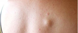
- skin neoplasms;
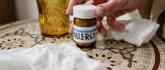
- fungal infections;
- hormonal imbalances;
- hypovitaminosis;
- inflammatory diseases;
- metabolic disorders in the body.
Excessive roughening of the skin is often explained by other ongoing diseases:
- psoriasis;
- ichthyosis;
- keratoderma;
- lichen;
- diabetes.
If we talk about foot hyperkeratosis, this form of pathology is often caused by improper gait or flat feet, excess body weight, and wearing tight and uncomfortable shoes.
Prevention
General preventive measures include:
- Compliance with sun exposure and solarium use.
- Strengthening the immune system.
- A rational diet that corresponds to energy costs.
- Prevention of trauma to the skin (wear comfortable, appropriately sized shoes, clothing made from natural fabrics).
- Maintain personal hygiene, correct (according to type) skin care.
- Regular use of medicinal/cosmetic products to moisturize and soften the skin and prevent peeling of keratinized plates.
- Avoid injury to the skin.
- Don't freeze.
- When working with any chemical reagents, avoid contact with skin (use special protective equipment).
- If you are prone to hyperkeratosis, regularly carry out hardware treatment methods and visit a podiatrist.
Symptoms of hyperkeratosis
The main symptom of the pathology (and its main characteristic) is thickening of the skin in the affected areas . Other symptoms may vary depending on the specific form of the disease.
With follicular hyperkeratosis, “goose bumps” appear, that is, dense reddish pimples with a spiky shape. They are located at the base of the hair follicles. Most often, all this is observed on the side or back of the thigh, buttocks, arms, and face. In rare cases, the patient may experience itching.
Plantar hyperkeratosis is characterized by roughening of the skin on the entire sole of the foot or only in the heel area. Calluses and bleeding cracks often occur, which causes discomfort and pain.
With the seborrheic form, characteristic pigmented formations appear, which over time lose color and become colorless plaques.
The symptoms of hyperkeratosis are more pronounced in adults; in children, the pathology is often similar to dermatitis. At the initial stages of development there are no symptoms.
Etiology and pathogenesis of keratosis
One of the significant factors in the development of keratosis is constant exposure to ultraviolet radiation . A special role is played by ultraviolet light B (UVB, 290–320 nm) and A (UVA, 320–400 nm) - it induces p53-dependent apoptosis of epidermal keratinocytes. These apoptotic keratinocytes are often called “sunburn cells” (SBCs) and can be detected by histological examination of epidermis that has been overexposed to sunlight or tanning bed lamps.
In the development of keratosis, the UV-induced mutation of the tumor suppressor gene TP53, which is related to the above-mentioned p53 protein, is of great importance. Basal keratinocytes with mutant TP53 do not undergo apoptosis, which allows them to pass on genetic abnormalities to new cells. Similar changes are found in squamous cell carcinoma and other malignant tumors.
Actinic keratoses may be promoted by a general suppression of the body's immune response. The risk group includes patients who have undergone organ or tissue transplantation and are taking immunosuppressants.
Some medications can act as photosensitizers, increasing the skin's sensitivity to ultraviolet light and promoting the development of keratoses cutis.
Microscopy of keratotic lesions usually reveals dysplastic changes in the basal layer of the epidermis, accompanied by parakeratosis and solar elastosis of varying severity. An increase in the size and pleomorphism of nuclei, hyperchromatosis, an increase in mitotic activity or an atypical nature of mitoses, and cytoplasmic pallor are recorded.
Dysplastic epidermal growths may extend deep into the papillary dermis and in some cases are difficult to distinguish from superficial invasive squamous cell carcinoma of the skin. Dysplastic epidermal changes, as a rule, do not affect skin appendages (sebaceous and sweat glands, hair, nails) ( Fig. 1 ).
Rice. 1. Histological features of keratosis (www.medscape.com)
In the epidermis, moderate hyperkeratosis is observed with dysplasia of basal keratinocytes and the formation of small outgrowths extending into the papillary dermis. The presence of solar elastosis is noticeable. Dysplastic changes primarily affect the interfollicular epidermis.
Stages of development
The following stages of disease development are distinguished:
- First . In some areas, the skin turns pale and becomes dry. No thickening of the stratum corneum has yet been observed.
- Second. When touching the skin, particles falling off are noticeable. Slight thickening is possible.
- Third . The skin peels off greatly and loses its smoothness, becoming rough and rough to the touch.
- Fourth. At this stage, multiple complications arise that lead to serious changes in the appearance of the epidermis. The thickening of the stratum corneum is visible to the naked eye.
Table of contents
- Etiology and pathogenesis
- Clinical features
- Principles of treatment
Keratosis (actinic keratosis, solar keratosis, actinic keratosis, actinic keratosis) is a precancerous skin condition that occurs as a result of a person’s prolonged exposure to the open sun.
In our company you can purchase the following equipment for the treatment of keratosis:
- M22 (Lumenis)
- Fraxel (Solta Medical)
- AcuPulse (Lumenis)
- UltraPulse (Lumenis)
Most often, actinic keratosis of the skin occurs in older people, but recently there has been a trend towards rejuvenation of this disease. According to statistics, the more time a person has spent in the open sun during his life and the more sunburns he has received, the higher the risk of developing keratoses as he ages.
Although a significant number of cases of squamous cell carcinoma are associated with actinic keratoses, the risk of the latter progressing to skin cancer is relatively low. Unfortunately, at present, no reliable histological criteria have been developed that could accurately predict which cases of actinic keratosis transform into skin cancer and which do not. According to various estimates, the probability of this event is in the range of 0.025–20%.
Diagnostics
Scleroderma can often be difficult to diagnose because it can be similar to many other diseases. Changes in skin thickness, the presence of certain antibodies in the blood, or early changes in blood vessels can be helpful in diagnosing the disease. If necessary, a biopsy can be taken to analyze the tissue, which also helps make the diagnosis.
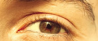
One useful test is nail capillary analysis. It is based on the early manifestation of scleroderma and is manifested by the disappearance of capillaries in the skin of the arms and legs. For this analysis, a microscope or magnifying glass is used and the skin in the nail area is viewed. For an accurate diagnosis, it is necessary to compare both laboratory research methods, clinical data, and medical history.
Principles of treatment of keratosis of the skin
As a measure to prevent the development of keratosis, it is recommended to constantly use sunscreen with SPF 30+. However, it is important to maintain a balance between photoprotection of the skin and possible vitamin D deficiency associated with a lack of sunlight. With active insolation or prolonged exposure to the street, the use of UV filters is justified, but if the patient spends most of the time indoors, there is usually no need for sunscreen cosmetics.
Some medications reduce the risk of actinic keratoses. For example, it has been found that with regular use of non-steroidal anti-inflammatory drugs, the incidence of keratoses is reduced. Medications can also be used to treat keratoses of the skin, but they should be prescribed in close consultation with dermatologists and other doctors. For example, the patient can be recommended 5-fluorocil, imiquimod, a topical combination of diclofenac and hyaluronic acid.
of chemical peels can be used .
Papules and skin horns can be eliminated using cryotherapy - local freezing with liquid nitrogen. This can also be done through surgery .
The problem of actinic keratosis is successfully solved by hardware methods , among which non-ablative fractional photothermolysis, as well as traditional and fractional laser ablation can be particularly highlighted. The latter combines well with photodynamic therapy - in this case, a fractional CO2 laser serves as a means of intradermal delivery of photosensitizers.