- Department of Aesthetic Medicine
Department of Aesthetic Medicine » - Injection cosmetology
Injection cosmetology »
- Services
Services "
- Regenerative therapy with autologous fibroblasts
Regenerative (or restorative) therapy with autologous fibroblasts is an amazing procedure that in 2010 moved from the field of experiment to the field of evidence-based medicine.
Regenerative therapy is a cell therapy that restores lost body functions through the transplantation of functionally active cells. The implanted cells release biologically active substances that have a healing and restorative effect.
The patient's own cultured fibroblasts are used for treatment
This unique method has received worldwide recognition as an effective and safe method. That is why regenerative therapy has been introduced into the daily practice of the RAMI Clinic.
Who is this technique for?
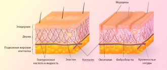
It is difficult to find a person in adulthood who would be satisfied with the condition of his skin. Over the years the number of active fibroblasts decreases , this leads to the appearance of wrinkles, drooping tissue of the eyelids or cheekbones.
Modern medicine can effectively replenish the deficiency of living skin cells. This is precisely what regenerative therapy with autologous fibroblasts is designed for, which is equally effective for both women and men .
In addition to correcting age-related changes, therapy with autologous fibroblasts is indicated for the treatment of post-acne scars, stretch marks, as well as during preoperative preparation and postoperative rehabilitation in plastic surgery.
Neural stem cells from fibroblasts
So, the production of neurons from fibroblasts is limited by the number of fibroblasts. In addition, the use of functional neurons for cell therapy is difficult - neurons form numerous processes, and they cannot be transplanted without cell death, since the processes are destroyed. What if we get neuronal stem cells rather than functional neurons? These cells have the potential to both divide and differentiate into the major neural cell types: neurons, glia, and oligodendroglia. Indeed, it has been shown that the expression of just one transcription factor, Sox2, makes it possible to obtain and then propagate in vitro neuronal stem cells, and upon their differentiation, neuronal cell types [18]. Apparently, obtaining neuronal stem cells is the future of cell therapy.
The uniqueness of regenerative therapy
Autologous dermal fibroblasts are the main human connective tissue cells that form the extracellular matrix. They play a leading role in the physiology of the skin: they regulate all metabolic processes, stimulate cell renewal, and participate in the healing processes of damage.
One of the main reasons for skin aging is a decrease in fibroblast activity. They slow down the synthesis of intercellular space, as a result, the moisture content decreases, the skin fades, and wrinkles appear.
Thanks to the introduction of autologous fibroblasts, tissues start their own metabolic processes, natural restoration and rejuvenation of the skin occurs.
Regenerative therapy technology is absolutely safe , does not cause allergic reactions or rejections, since only natural ingredients are used - the patient’s own cells .
The effectiveness of the regenerative therapy method
The effectiveness of the technique has been proven by numerous clinical studies in the USA, Europe and Russia.
Specialists from the Ural State Medical Academy and the Institute of Medical Cell Technologies observed 45 patients aged 43 to 60 years who received autologous fibroblast transplants for two years.
As a result , it was revealed that after the procedure, patients’ fine wrinkles disappeared and deep wrinkles were corrected, skin turgor and elasticity increased, and its thickness increased. The effect is increasing in nature, reaches a maximum after 15 months (after the introduction of cells) and persists for at least two years. There were no side effects after the procedure during the observation period.
In 2012, at the RAMI multidisciplinary clinic, modern technology for non-surgical skin rejuvenation based on cell therapy was introduced into everyday practice.
This was preceded by the multifaceted work of our clinic staff :
- studying scientific publications on the use of cellular technologies in cosmetology, visiting international forums;
- participation in experimental work on cell cultivation, consultations with foreign and Russian colleagues;
- search for partners licensed to conduct biotechnological work;
- formation of appropriate material and technical base and human resources of specialists.
Competition "bio/mol/text"-2013
This article was submitted to the competition of popular science works “bio/mol/text”-2013 in the category “Best News Message”.
The competition is sponsored by the visionary Thermo Fisher Scientific. The sponsor of the People's Choice Award is Helicon.
Cell therapy is one of the most promising areas of medicine. Replacing damaged or dysfunctional cells could be a way to treat diseases that are currently considered incurable. In general, cell therapy can be divided into several areas: transplantation of progenitor cells, adult stem cells, transplantation of differentiated cells ready to perform their functions, and transplantation of genetically modified cells with corrected defects. Currently, the use of cell therapy in practice is limited to blood transfusions and bone marrow transplants.
For the widespread use of cell therapy in the treatment of diseases, several problems must be solved. First, the introduction of foreign cellular material causes immunological rejection. During blood transfusion, the compatibility of the blood type and Rh factor of the donor and the patient is monitored. When transplanting organs and bone marrow, it is necessary to take into account the coincidence of antigens of the major histocompatibility complex, which significantly limits the ability to find a donor in time. And even when using “compatible” material, recipients are forced to take immunosuppressants throughout their lives.
Secondly, the ability to obtain donor cells is physically limited - only certain cell types are available - for example, bone marrow cells, but the available number of even these cells may not be sufficient for treatment. If we talk about neurons, hepatocytes and other specialized cells, then it is almost impossible to obtain them. Thirdly, differentiated cells of an adult organism are most often no longer capable of division, are in their cellular niche and are unsuitable for transplantation.
Nervous system diseases associated with the dysfunction and death of neurons, such as Alzheimer's and Parkinson's diseases, currently have no cure. Multiple sclerosis, a disease associated with demyelination of axons in the central and peripheral nervous system, is also incurable. At the same time, it is known that, for example, a sign of Parkinson’s disease is the death of dopaminergic neurons in the substantia nigra of the midbrain, and in multiple sclerosis, oligodendrocytes stop producing myelin. Cell therapy and transplantation of functional neural cells may be a promising approach to treat such diseases.
Below we will discuss various methods for obtaining neural cells for cell therapy, prospects for application and existing problems.
How is regenerative therapy treated?
Regenerative therapy with autologous fibroblasts involves 2 stages: clinical and laboratory (biotechnological).
Clinical stage
Takes place in the multidisciplinary clinic “RAMI”.
First, a comprehensive examination of the patient is carried out , indications for the use of this treatment method are established, possible contraindications are identified, and the optimal regimen for administering the drug is determined.
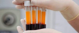
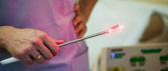
Then specialists from the RAMI clinic take a flap of skin from the behind-the-ear area measuring 0.6*0.6 cm. The resulting biopsy is transported to a specialized laboratory for cell processing (cultivation).
Laboratory stage
It is carried out in a specialized institution that has a license to conduct biotechnological work, isolate, cultivate and store cell cultures. The fibroblast culture obtained in the laboratory is tested for stability, oncological and infectious safety. Each drug that contains a therapeutic dose of cells is issued a corresponding passport .
For intradermal injection into the patient's problem area, a certain therapeutic dose of fibroblasts (at least 1 million cells) is required, which is determined by the doctor.
The collection of a skin flap, its transportation, and intradermal introduction of cells are carried out under conditions of maximum sterility.
The time for culturing cells and forming a fibroblast culture in a specialized laboratory is 25 - 29 days. The resulting fibroblast culture is frozen in liquid nitrogen and stored for the required time in compliance with all requirements.
Next, a preparation of living fibroblasts is prepared from the resulting culture in the form of a suspension, 1 ml of which contains at least 1 million functioning fibroblasts. The time required to prepare the drug is 5 - 7 days.
New skin: can't we stand on the price? Part 2. Where fibroblasts grow
Having given a piece of skin for the production of new fibroblasts, Yana had to wait for them to grow. But I couldn’t resist and went to the laboratory to visit them. A month ago, I announced that in an attempt to preserve and increase youth and unearthly beauty, I was going to extreme measures :) - I was putting myself in the hands of SPRS therapy. In a nutshell, the essence of it is that they first take a skin biopsy from you, then grow new fibroblasts from it, then inject them into you - and you, like in a fairy tale, emerge from this rejuvenating cauldron with a brand new face. (For more details, see the links. You can read the beginning of the story here.)
Usually, all SPRS therapy patients, having parted with a piece of skin, wait patiently for a couple of months until they receive a call, say that everything is ready, and schedule a day for injections. But I’m a special patient - distinguished by nasty curiosity. Therefore, I agreed with one of the creators of the SPRS therapy method (and expert consultant of our blog) Vadim Zorin that I would come to visit my fibroblasts. I will find out, so to speak, how they behave. Are they being naughty or capricious? And then you never know. Still not strangers. Then blush for them.
Moreover, it turned out so fortunately that my fibroblasts grow in close proximity to me - on Gubkin Street. This is where the Human Stem Cell Institute (HSCI) is located, one of the divisions of which is SPRS therapy.
While I was visiting my fibroblasts, it started to rain. I took this as a good sign: everything grows well in the rain)
The laboratory and production complex of SPRS therapy is literally behind seven seals and ten doors, as it should be either in fairy tales or science fiction novels. You can't get through without a guide. My Stalker was a charming girl (and my namesake) Yana.
To get inside door N1, you had to say a secret word, dial a secret code, bow to the east three times and test yourself for purity of thoughts
Then I had to turn into either a penguin or an astronaut. Put on these sterile paper shoe covers...
This is a staged shot that allows the author to demonstrate Chanel sandals :) Actually, shoe covers had to be put on both feet, of course
...here is a sterile paper spacesuit...
Fits into size S. Not a big deal, but nice.
... and go through purgatory - a transitional vestibule. There we also had to wash our hands and put on a mask. I have been in a wide variety of sterile places - for example, in the labor room, where I watched babies being born, and in the operating room, where I assisted in mammoplasty and liposuction and watched how a circumferential lift was done. But nowhere was such a degree of sterility required of me. And here I literally miraculously escaped brain disinfection :)
Vadim Zorin met me in purgatory. He was strict - he ordered her to put her bangs under her hood. However, he is always strict - this seems to be his usual state.
Door N33. It is already clear that my fibroblasts are under reliable protection. They are protected like some kind of state secret
The “place of power” of fibroblasts turned out to be a rather small room - about like my bedroom, 20-25 sq.m. But apparently, the cost of these square meters greatly exceeds mine. For example, I have been unable to install an air conditioner at home for several summers in a row - but here there are special filters for air purification, each for the price of a cast-iron bridge. And here is such a device, vaguely similar to the ZIL refrigerator.
It seems like we have only one such incubator in the whole country. Just like the ZIL refrigerator - in my opinion, it was left only with my mother. She doesn’t want to part with him - she says he’s dear to her like a mausoleum of her youth :)
But it turned out that this is not a refrigerator, but a CO2 incubator, where a constant temperature of +37 degrees is maintained (the same as the temperature of the human body), and where fibroblasts grow in an atmosphere that is a mixture of oxygen and 5% carbon dioxide.
Fibroblasts are located in this incubator in special containers with a special solution. The containers are labeled with the names of the fibroblast hosts. Of course, I’m interested in how many people who don’t want to meekly wait for old age and are willing to pay from 280,000 rubles for new skin. But this trade secret is a great one. Who will tell me this? True, Dr. Svetlana Shokolova, who took a biopsy from behind my ear at the RayLife clinic, said that there are many patients, and there are those who come to her “for transplantation” not only from Khabarovsk and Yekaterinburg, but also from France with periodically every two to three years.
But, in any case, I'm not alone. A review of the contents of the ZIL refrigerator incubator once again confirmed this.
The containers where fibroblasts multiply are different. Some have one compartment, some have three. Fibroblasts do not float in a nutrient medium, but live on the bottom. As they multiply, they are moved - much like a family, as they add more offspring, moves to a larger living space. Only this move is carried out with a lot more formalities.
Naturally, I asked Zorin to show me my personal fibroblasts. In the end, it was them that I came to visit. Zorin found them easily - all containers were labeled.
On my container there was a label: “Zubtsova. Super."
The triumph of modern science as it is. A man holds his own cells in his hands. An exciting moment, I must tell you
After reading the word “super,” I was as happy as a mother at a parent-teacher meeting when it was suddenly announced that her child had suddenly received an “5” on his annual physics test. (A feeling that I have never experienced in my years of motherhood:)))
A moment of pride in your own fibroblasts
Of course, I immediately decided that my fibroblasts were somehow especially successful and cute, somehow reproduced well and did not cry in captivity, since they were “super”.
Ooops.
It turned out that “super” has nothing to do with their behavior and is not an evaluative characteristic at all, but, let’s say, the name of one of the stages of their development.
Zorin showed how fibroblasts lie on the bottom of the container. Their bed is called a “mattress”. I pretended that I saw something there, although in fact I saw absolutely nothing.
Then Zorin took the container and placed it under a microscope connected to a monitor. The monitor displayed something reminiscent of a painting by an unknown primitivist artist: “Someone scattered black, long, long grains of rice on a gray, gray table.”
These “grains of rice” are fibroblasts, I guessed. Here, on this mattress, there are still few of them. After some time, they will multiply and occupy the entire surface. Then they will be moved to a two-level container.
In this container, fibroblasts are just beginning to divide; quite rare single representatives are visible on the screen.
Then Zorin put another container under the microscope - with fibroblasts, almost ready for replanting. The screen displayed something that looked like the fingerprint of a space alien. “See these numerous schools of fish? These are already entire “populations” of fibroblasts,” Zorin explained. And I was once again convinced of how differently people see the world. Some have an alien finger, others have schools of fish :)
What do you think - a joint or an alien finger? Or maybe there are other ways to read this painting?
“And the white blots on the screen are cells that are currently dividing. In order to divide, the fibroblast needs to turn into such a “blot”, from which another joint will then bud.” He explained everything to me like a fool, trying to avoid scientific terms. And he did the right thing. Otherwise I definitely wouldn’t have understood anything.
In addition to Zorin and me, fibroblasts, a ZIL refrigerator and microscopes, in the laboratory there is another employee of SPRS therapy - and two objects that vaguely reminded me of counters with frozen semi-finished products in supermarkets.
“Laminar flow box” is the name of this “counter”
It turned out that these “counters” are called laminar flow cabinets. A stream of sterile air circulates inside such a cabinet. Only under such conditions is it possible to carry out any manipulations with fibroblasts “in open space,” that is, in the air.
In appearance, the solution—the nutrient medium in which fibroblasts live and multiply—looks like highly diluted blood. In fact, there are 300 components in this solution, mainly amino acids and vitamins. And there is not a drop of blood.
But it turns out there’s a lot of trouble with them. For one procedure for introducing fibroblasts into one area (for example, into the skin of the face), there should be at least 60-80 million of them. And each of these 80 million should be in full combat readiness by the time of replantation - that is, ready to produce the correct collagen and elastin. That’s why they are treated here like little children in an elite kindergarten. They constantly change their nutritional medium, transfer them from one mattress to another as they reproduce, in general, they keep a vigil and don’t take their eyes off them. And all this is carried out in these very laminar flow cabinets.
And, to the question of why the SPRS therapy procedure costs the money it costs. The cost of 1 square meter of the laboratory where we are now is 10,000 euros. Not counting the equipment. Just the cost of the air filtration and purification system that is installed here. I can’t even imagine how much the equipment costs. But there are also reagents, substances, the work of professional technologists, in the end.
The nutrient medium is changed once every 3-4 days. And once every 7 days, the cages are transferred from one mattress to another.
Before becoming a nutrient medium for fibroblasts, its components are stored in bottles like these. Each one has an expiration date written on it - and, unlike many other cases known to us, it is not violated even for an hour.
It is important to correctly determine the date when fibroblasts reach the peak of their development and are ready for “replanting”. The patient is scheduled for the procedure on this day. If he suddenly cannot come for some reason, you need to notify him in advance so that the laboratory has time to carry out special manipulations to preserve the cells. But fibroblasts cannot sit on their mattresses and wait forever. After a certain point, they must either be put into use or frozen.
There is another microscope in the laboratory through which you can observe the well-being of fibroblasts. We admire mine once again. Yes, for control. Just in case.
I realized that they were cute after all. Obviously more beautiful than, say, patient Ivanov or patient Sidorov.
After which we leave the laboratory, take off our paper spacesuits (more precisely, I took them off, and Zorin simply changed into civilian clothes) and go to another building, where the fibroblast cryogenic storage facility is located.
A door is like a door. It looks like the door to a school chemistry or physics classroom.
There, behind this door, are two fairly large halls where there are containers with liquid nitrogen. Inside each one the temperature is -185 - -196 degrees Celsius.
And these refrigerators seemed to me like top-loading washing machines.
Inside these “washing machines” there is permafrost.
Zorin put on gloves and a mask, opened one of the “washing machines” and immediately found himself enveloped in icy nitrogen vapor.
Note to the hostess: ice steam burns hotter than hot steam.
And I remembered that the air swirled in approximately the same way in Finnish cryosaunas. This is such a popular thing in Finland - cold treatment, cryotherapy. Many spas there have cryosaunas. This seemed terribly funny to me, considering that I was there in the winter, and outside, without any cryo-spa, it was minus 35. It was worth building something special, if you go out into the forest naked - and here you have cryotherapy in life-size, I thought. And the Finns explained to me that if you go into this refrigerator and wander around there for a couple of minutes, cellulite and psoriasis will disappear. You must enter naked (well, in a swimsuit), felt boots, a hat and mittens. What a spectacle it is. But the impressions are unforgettable.
While I was reminiscing, Zorin took out of the refrigerator a metal shelf covered with a layer of ice, a la Ikea, and showed me in what form the fibroblasts were actually stored there.
By the way, each test tube is frozen gradually, lowering the temperature by 1 degree per minute. So this is also a whole ritual and art.
They are stored in cryovials, to which a cryopreservative is added so that the cells can more easily withstand permafrost conditions and do not burst. This preservative is something like that special protein that is produced in the body of frogs. After all, it is known that frogs can freeze through and through, and then defrost - and feel great. Whereas a bear, for example, in a similar situation would be torn apart from the inside.
In one such test tube, you can see the youth of someone’s skin 100 decades into the future. I can't wrap my head around it.
To create such a bank of fibroblasts, all you need is the same one piece of skin that was taken from me. Cells can be stored frozen in cryostorage for as long as desired—more precisely, as long as their owner is willing to pay for such a “cryo-spa.” By the way, it’s not too expensive - 6,000 rubles. in year. But before the next “replantation” you do not need to take a skin biopsy again and carry out a series of manipulations with it. And, by the way, this bank helps out a lot if some kind of emergency happens, for example, a fire, and you get serious burns. Then these fibroblasts are extracted, thawed, a cell preparation is quickly prepared from them - and doctors use it to restore the integrity of the skin.
As we left the cryogenic storage facility, I noticed a pile of mittens - apparently they belonged to the maintenance staff.
God have mercy on polar explorers!))
It reminded me of a school locker room, where in winter the mittens lost by first-graders during the reporting period are piled up - and such a household association somehow brought me back to earth. Because until then it still seemed to me that, although I was already without a spacesuit, I was still in space, in the future, in the novel by the Strugatsky brothers, in general, anywhere, just not in Moscow in 2014.
And then I realized where I was, what I was, why I was, I grouped up and asked Zorin a question that had worried me from the very beginning of this whole story. Because why freeze and store newborn stem cells taken from the umbilical cord, I can understand that. You can fall back on them, as a source of rejuvenation, throughout your life. Why store fibroblasts of a young maiden - too. In 20 years she will no longer be young, but they will be. But mine?.. I'm no longer a child. And, alas, not even a teenager. And my cells correspond to my passport more than, for example, my cheerful appearance - because the entire global beauty industry works for it, and, alas, only my wrong lifestyle works for them. And it would be more accurate to say not “on them”, but “against them”. In my opinion, even now my fibroblasts are of dubious value. And in ten years?..
Zorin listens to me patiently, but in his eyes I see that he has been asked this question once before. And that he had long ago come to terms with what they would ask in the hundred and tenth millennium. “No,” he says, “for SPRS therapy it doesn’t matter whether we take fibroblasts from a 20-year-old, a 40-year-old or a 60-year-old. No matter who we take them from, natural selection of cells occurs in the process, and as a result, only the most active and capable ones, capable of dividing and synthesizing collagen and other important components, remain for reproduction. All others are removed from the process. We did a lot of research. Cultured fibroblasts taken from 18-year-old and 82-year-old people were compared. There was no difference between these fibroblasts: they produced collagen and elastin at the same high levels.”
He explains all this to me casually, like a teacher explaining the multiplication table to a student for the hundredth time. And he gives several books and magazines with his articles on this topic.
I leave loaded with scientific literature and with serious intentions to get into it all. And still I really feel like a guest from the past, caught in a time machine in some next century.
In a couple of weeks I will be given fibroblasts. Will a new life begin?..
In the next parts of the series “New Skin. Can’t we stand behind the price?”:
— report on the introduction of fibroblasts;
— interviews with people who have been injecting themselves with fibroblasts regularly for a long time;
— a story about what this genetic skin passport is and whether it is worth pinning serious hopes on it;
— stories about what a patient who has undergone SPRS therapy feels and looks like one month, two months, six months and a year after the procedure.
Photo: Ksenia Nikolaeva (website - www.xenya.ru, facebook page)
The process of introducing cells into a problem area
The schedule for introducing cells into the problem area of the skin depends on:
- the time required to obtain a cell culture;
- therapeutic dose of a living cell drug;
- individual characteristics of the patient's skin;
- from choosing the optimal treatment regimen recommended by the doctor.
Cell injection area
The area into which fibroblasts are injected intradermally is determined jointly by the doctor and the patient. As a rule, problem areas of the face are selected - around the eyes, nasolabial folds. The cell preparation can also be injected into the neck and décolleté area.
Of particular interest is the use of autologous fibroblasts for rejuvenating the skin of the dorsum of the hands. Autologous fibroblasts can be injected into the area of scars, stretch marks, and wrinkles.
Depending on the depth and severity of skin changes, a papular or tunnel method of administering the cell preparation is used.
Live cell injection procedure
- the procedure is performed under local anesthesia. The doctor injects a drug enriched with fibroblasts into the patient’s problem area with a thin needle;
- depending on the problem, several sessions may be required;
- the procedure is performed under local anesthesia. Using a thin needle, the doctor injects a drug enriched with fibroblasts into the patient’s problem area;
- After the procedure, slight redness and swelling may occur, which will disappear within 2 - 3 hours.
When active fibroblasts are introduced into the skin, the natural processes of restoration of the dermis are launched. The effect of the procedure begins to appear 6-8 hours after the session and increases over time. 8 - 9 months after the course of treatment, a persistent positive result is observed.
Result after regenerative therapy
After the introduction of cultured fibroblasts, the number of dermal cells in the treated area increases, the synthesis of its own collagen increases, microcirculation is restored, metabolic processes and blood circulation are stimulated.
The high efficiency of the method is due to the ability of implanted living cells to produce biologically active substances characteristic of young skin for a long time (8-9 months): its strength and elasticity increases, and a lifting effect occurs.
The result of implantation of living cells: wrinkles, scars, and stretch marks disappear. The dermis is completely renewed, healthy skin color , its turgor increases, the skin becomes velvety , dryness and flaking disappear.
Difference from other methods
The main difference from other methods is the drug used. The patient's skin is restored using its own cells , which are cultured in the laboratory.
During the procedure, living cells are transplanted onto the skin, which dramatically accelerate the skin regeneration mechanism due to their production of growth factors and components of the dermal framework. The method not only corrects visible skin defects , but also restores it at the microtexture level.
Modern cosmetological techniques (antioxidants, hydrants, elastic stimulants, biostimulants, capillary protectors, etc.) have a certain rejuvenating effect, but the principle of action is based on correcting the effects of aging. Cell replacement therapy solves the problem of skin aging on a fundamentally different level: the process of reducing the reserves of one’s own skin cells stops. This means that the aging process in the treated area is not only stopped, but even reversed.
Regenerative therapy treats age-related skin changes when other methods only correct their consequences.
Cell fate and how to change it
The entire variety of cell types in normal development—neurons, skin cells, blood cells, liver cells, and others—is formed as a result of the division and differentiation of a single cell—the zygote. A zygote is formed by the fusion of gametes—an oocyte and a sperm. Then, as a result of cell division, a group of cells is formed - a morula. Next, a cavity is formed - the blastocoel; this stage of embryonic development is called blastocyst. The outer cells - the trophoblast - will participate in implantation and formation of the placenta, and all types of embryonic cells will develop from the inner cell mass. Embryonic development can be thought of as a tree, based on a single cell (zygote), and the branches extending from the trunk symbolize the sequential differentiation of cells. The analogy with the stem is also convenient because embryonic stem (ES) cells, which will be discussed further, are obtained from the cells of the inner cell mass. The cells of the inner cell mass are undifferentiated: they are capable of becoming any type of cell. Each branching of the tree is a choice of fate in the process of differentiation, specialization of cells and narrowing of the spectrum of differentiation. First, there is a division into three germ layers - endoderm, mesoderm and ectoderm. We can additionally distinguish the germinal path and future gametes. Very schematically, further development can be represented as the formation of cells of such organs as kidneys, lungs, stomach from the endoderm; muscles, heart, spleen, blood - from the mesoderm; nervous system and epidermis - from ectoderm. The ability to divide and differentiate into cells derived from the three germ layers is called pluripotency. The developmental program is carried out through the sequential activation of various groups of transcription factors that regulate the expression of many genes. Gene expression is influenced by both internal factors and external signals, such as those received from surrounding cells. Adult stem cells maintain the vital functions of organs and tissues. The different types of stem cells have already been discussed on the “biomolecule”: “Trunk and branches: stem cells” [1].
In experimental systems and in some cases in normal development, changes in cell fate are possible (Fig. 1). If differentiation is the transformation of an undifferentiated cell into a differentiated one, then the reverse process is called dedifferentiation. The transformation of one cell precursor into another is transdetermination. For example, during the process of hematopoiesis, hematopoietic stem cells differentiate into precursors of the lymphoid series (lymphocytes, etc.) and the myeloid series (granulocytes, macrophages, erythrocytes, etc.). It turned out that in an experimental system it is possible to induce transdetermination - using the expression of transcription factors, to change the fate of the precursors of the myeloid series so that they differentiate into the precursors of granulocytes and macrophages, or, in another experiment, into the precursors of erythrocytes [2]. Finally, direct differentiation has recently been demonstrated—embryonic stem cells can be directly differentiated into neurons through the expression of specific transcription factors. This differentiation is called direct because the stages of differentiation required in normal development are absent in this case. Finally, the transformation of a differentiated cell of one type into another without going through the stages of dedifferentiation is transdifferentiation.
Figure 1. Different pathways for changing cell fate. Normal development is the differentiation of a totipotent zygote into various types of cells with a gradual narrowing of the potential for differentiation. In an experiment, you can change the fate of a cell—induce dedifferentiation, transdetermination, or direct differentiation.
All of the above methods for changing cell fate are potentially suitable for use in cell therapy. The possibilities of using these approaches will be discussed below, with an emphasis on obtaining neural cells and using a relatively new direction in research, transdifferentiation.
The following sources were used in preparing the material:
- “30 years of experience in the development and application of cell technologies in clinical practice.” V.P. Tumanov, D.A. Zhakota, N.S., Korchagina. Scientific and practical journal “Plastic surgery and cosmetology”, 2012 (3), Moscow, Russia.
- Isaev AA, Prikhodko AV, Zorin VL, etc. Medical technology “Collection, transportation, isolation, cultivation, cryopreservation, storage and use of autologous fibroblasts for the correction of age-related and scarring changes in the skin.” FS No. 2009/308 dated July 21, 2010, Moscow, Russia.
- “The use of autologous dermal fibroblasts to correct age-related changes in facial skin. Results of one-year research.” Authors: V. Zorin, A. Zorina, V. Cherkasov et al. Journal “Aesthetic Medicine”, volume XI No. 2, 2012, Moscow, Russia.
- “Qualitative and quantitative assessment of the condition of facial skin after the use of autologous dermal fibroblasts.” Authors: V. L. Zorin, A. I. Zorina, V. R. Cherkasov, P. B. et al. Journal “Bulletin of Aesthetic Medicine”, volume 10, No. 2, 2011, Moscow, Russia.
Perhaps you will find it useful:
- Post-traumatic, post-burn regeneration of the skin
If you are really looking for your doctor...