There are a large number of types of skin formations (both benign and malignant). They often do not pose a risk to your health, but they can cause physical or psychological discomfort, especially in areas of pressure, friction, areas of skin at risk of injury, and on visible parts of the body. You also need to know that some skin formations may be prone to discoloration, growth, and malignancy. Today we will talk about the types of skin formations and their possible changes, the appearance of which you need to be wary of. We will also look at the most delicate and gentle way to remove tumors.
What research is done before removing skin tumors?
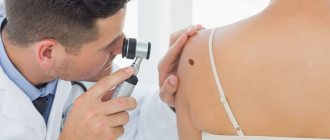
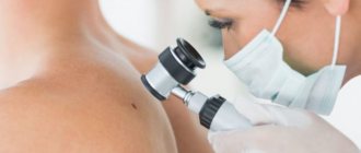
The main optical device used by a dermatologist to examine moles and other skin growths before their removal is a dermatoscope. A dermatoscope consists of a magnifying lens and a light source to improve the visibility of subsurface structures of the skin.
The examination procedure using a dermatoscope is called dermatoscopy. This diagnostic method allows an experienced specialist to visually assess and diagnose the nature of a skin tumor under tens of times magnification.
How much does it cost?
Removal of 1 milia (grass, wen) in the eye area
950 rub.
Removal of 2 skin lesions on a wide base
(removal 8700, histology 2300)
11000 rub.
Removal of 3 skin lesions on a wide base
(removal 10400, histology 4600)
15000 rub.
If you doubt the price, find out the exact cost in advance. To do this, take a photo of the tumor against the background of a centimeter scale ruler and send it to my email address
Dmitry Beinusov: examination + consultation + surgery
- Dermato-oncologist, Candidate of Medical Sciences.
- Postgraduate studies at the Oncology Research Institute named after. N. N. Petrova.
- Specialization: skin neoplasms.
- Member of the European Association of Surgical Oncologists (ESSO).
- Author of 10 scientific articles and holder of 1 patent.
- Permanent consultant of the portal Health Mail.ru
- Teacher of advanced training cycle in dermato-oncology
What is the best method for removing skin tumors?
Our clinic's arsenal includes almost all methods for removing any benign skin tumors, except for surgical and chemical destruction: laser removal, radio wave removal - radiosurgery, diathermocoagulation - plasma coagulation and cryodestruction - removal with liquid nitrogen.
In each individual case, the question of choosing a removal method depends on the nature of the tumor, its size, location, and concomitant pathologies. Therefore, the concept of the best removal method simply does not exist.
Symptoms and causes, what to look for
Symptoms of a soft tissue tumor can vary widely. Most often, the clinical signs of benign neoplasms are the appearance of a painless but constantly growing lump. The sizes of formations can be very different, it all depends on the place of their appearance. Typically, tumors that form on the head, neck or upper extremities are small in size due to early detection. Whereas formations in the abdominal cavity and thighs can reach significant sizes.
It is worth noting that there are no 100% signs that could help distinguish a benign from a malignant formation. Therefore, during a clinical examination of benign soft tissue formations, there is always the possibility of finding a malignant tumor.
In most cases, the appearance of tumors on the skin has no reason. Most often, such tumors arise spontaneously. In some cases, the cause of benign tumors is a genetic predisposition.
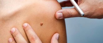
How can a tumor be removed without leaving a trace on the skin?
All superficial neoplasms - papillomas, keratomas, etc., as well as small formations, are most often removed without scars or marks on the skin. After removal of large formations, traces of hypopigmentation or barely noticeable whitish scars may remain.
In any case, it is very important when removing! clearly assess the line separating the difference between the aesthetic result of the procedure and its effectiveness. This means that the wider and deeper the tumor is removed, the lower the likelihood of relapse. Conversely, minimal superficial removal is always an ideal cosmetic result, rapid epithelization, absence of a scar, but a high risk of developing recurrent neoplasm at this site. The experience of our doctors, mandatory diagnostics and the availability of a wide range of equipment allow our specialists to maintain a competent balance between removing the tumor in full and obtaining the best possible cosmetic result of the procedure.
Complications of benign neoplasms
Even if a benign neoplasm is not accompanied by unpleasant symptoms such as pain, growth or suppuration, it is necessary to be regularly examined by a dermatologist. The following types of formations are prone to malignancy:
- moles and nevi
- papillomas (in the form of a growth on the leg)
- condylomas (warts in the genital area)
- dermatofibroma (in rare cases)
Frequent damage and infection in the wound leads to inflammatory processes, which may involve surrounding tissues. Inflammation is accompanied by pain, if an area of exposed skin is affected, this is accompanied by severe psychological discomfort.
Timely removal of skin lesions prevents the risks of complications and reduces the volume of surgery.
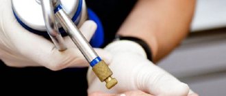
What are the advantages of the radio wave method compared to other removal methods?
The radio wave method for removing skin tumors is the most non-traumatic procedure, which is based on the use of high-frequency waves (radio waves) on the Surgitron apparatus, USA. This method is used even if it is necessary to remove an element located in a hard-to-reach place (oral mucosa, eyelids).
The advantages of the radio wave method include:
- complete absence of pain during the procedure;
- no bleeding after removal;
- no trace remains at the site of removal;
- no risk of secondary infection;
- healthy areas remain untouched;
- fast healing process;
- The method is universal - you can remove warts, papillomas and moles.
One of the important differences between radiosurgery and laser and other destructive techniques is the possibility of performing a histological examination of removed tissue when using the radio wave method of removal.
Indications for histological examination
Histological examination differentiates a benign process from a malignant one. For histological analysis, fragments of a tumor or mole taken during removal are used. Histological analysis consists of examining thin sections of pathological tissue using an electron microscope. If histological results show signs of a malignant process, the skin in the area of the removed tumor is excised more widely.
All tumors removed surgically or excised by other methods can be subjected to histological examination. With laser destruction, tumor tissue is destroyed, and histological analysis becomes impossible.
What happens after removal?
During the first day after the procedure, a dark brown scab (crust) will form at the site of the removed tumor. Healing takes from 2-3 days to 2-3 weeks, it all depends on the size of the tumor bed. After final epithelization, the crust will be torn off on its own. Within 1-3 months, the area of skin at the site of removal has a bright pink color, then it will acquire a normal skin color. If you follow all the doctor’s recommendations and properly care for the skin at the site of removal, no marks or scars will form.
Removal by radiosurgical method - indications and contraindications
Recommendations: Indications for radiosurgery:
- warts on the sole;
- papillomas;
- Nevi (moles);
- spider veins;
- keratoses;
- vulgar warts;
- Hemangiomas
- Skin basal cell carcinomas
- Ingrown nail
- removal of granulation tissue after surgery;
Attention: Contraindications:
- acute inflammatory processes;
- allergy;
- presence of viral infections;
- feverish condition;
- severe liver and kidney diseases;
- diabetes;
- period of bearing a child;
- herpes
Are there any special recommendations for skin care after the removal procedure?
We issue the following recommendations in the form of a reminder to our patients:
- During the first 24 hours, do not wet the removal site.
- Treat the wound with Fukortsin solution 2 times a day for 3-5 days or Baneocin powder.
- Men: do not shave in the area of removal for 3-4 days.
- Women: do not apply cream or decorative cosmetics to the removal area for 3-4 days.
- You cannot pick or remove the crust yourself!!! in order to reduce the likelihood of atrophic/keloid scars, hypo- and hyperpigmentation.
- In cases of removal of large tumors, after the crust is rejected, apply Levomekol ointment locally 1-2 times a day for a month.
- Before going outside, be sure to use sunscreen on the face and open areas of the body with an SPF of at least 50, especially on sunny days (to prevent pigmentation of the treated areas).
Treatment of benign non-pigmented skin lesions
Papilloma (papillary polyp, papillary fibroepithelioma) is a benign tumor developing from the epithelium; has the appearance of a papillary growth protruding above the surface of the surrounding tissue. The process characterized by the formation of multiple papillomas is called papillomatosis. Papillomas are found on the skin, mucous membrane of the mouth, nose, paranasal sinuses, pharynx, esophagus, larynx, trachea and bronchi, renal pelvis, ureters and bladder, as well as on the labia, vagina, and cervix.
In most cases, papillomas are of a viral nature , i.e. The proliferation of skin tissue is caused by a special virus (HPV - human papillomavirus), transmitted from person to person through direct contact (contact-household and sexual transmission of infection). Microscopically, papilloma consists of connective tissue stroma and epithelium, which may vary depending on the location of the formation. Depending on the amount of connective tissue, papilloma can be soft or dense.
Clinically, papilloma is usually a circumscribed tumor, up to 1-2 cm in diameter (sometimes large), dense or soft to the touch, on a thin long or short stalk, less often on a broad base . The surface of the papilloma is uneven, fine- or coarse-grained, reminiscent of cauliflower or cockscomb. Skin papillomas can have different colors - from white to dirty brown (depending on the blood supply to the vessels and pigment content). Skin papillomas usually do not cause any particular complaints in patients except for a cosmetic defect. Papillomas of other localizations can lead to various complications.
The indication for papilloma removal is the elimination of cosmetic defects , as well as the localization of the tumor, which can lead to functional disorders of the affected organ, trauma to the papilloma and repeated bleeding, inflammation, which in turn is associated with a high risk of malignancy.
All types of local treatment are aimed at removing papilloma and atypically changed epithelium, depending on their location. Various types of chemical coagulations, applications of cytostatic drugs and physical-surgical methods, surgical removal are used. Recently, immunological treatment methods have become widespread - using α, γ-interferons, as well as combined procedures, for example, the combined use of different treatment methods (cryotherapy, laser removal, electrocoagulation, diathermocoagulation).
Today, there are several methods for surgical removal of these formations:
- Chemical coagulation : for this purpose, the application of the drug Solcoderm is used, which is a mixture of organic and inorganic acids that cause the destruction of pathologically altered epithelium. The method is characterized by a slightly painful procedure; it is used to remove single skin papillomas, but may leave scars on the skin after use (difficult to control destructive effects).
- Cryodestruction is the destruction of pathologically altered epithelium by exposure to low temperatures (-96 degrees). A very effective, fast and painless method of removal, effective for single small papillomas without the risk of malignancy, because the method does not provide deep destruction.
- Electrocoagulation (DEC) is the destruction of pathologically altered epithelium by exposure to high temperatures (burning). This is a rather painful technique that gives the highest number of complications, accompanied by prolonged healing and poor cosmetic effect (scar formation). However, it is characterized by high radicalism (total destruction of the affected tissue) and is used when malignancy is suspected.
- Surgical laser is the preferred method of eliminating papillomavirus infection. It is characterized by slight pain, the ability to sting all changed areas to the required depth under eye control, and the absence of scars.
- Radiosurgery (radio knife, Surgitron apparatus) - destruction of the epithelium under the influence of electromagnetic waves. A painless, fast and reliable technique that allows you to cut out a section of tissue with virtually no bleeding and take it for histological examination.
- Scalpel - surgical excision of the tumor along with a section of healthy tissue under local anesthesia, followed by the application of a cosmetic suture. The most radical and reliable way. Allows for histological examination of the formation, which is especially important if malignancy is suspected (chronically injured papilloma).
However, it should be remembered that after removal of the papilloma, the virus may remain in the cells of the surrounding tissues, and papillomas may appear again. In these cases, it is necessary to consult an infectious disease specialist and decide on systemic therapy.
Senile keratoma
Senile keratoma (seborrheic keratoma, senile wart) is a brown or bronze formation with an uneven bumpy surface and clear boundaries, convex, as if glued to the skin. Senile keratomas are benign and occur mainly in older people. They are located on the face or body, are usually numerous and range in size from a few millimeters to several centimeters. If the tumor is damaged (most often by tight clothing), crusts may form on its surface and bleeding may occur. Treatment: cryodestruction with liquid nitrogen, surgical excision of the keratoma. Senile keratoma does not require treatment if it does not cause cosmetic discomfort and is not injured by clothing .
Solar keratosis
Solar keratosis - actinic, or senile keratosis, is classified as a precancerous disease . The disease begins with the appearance of scaly plaques, often accompanied by varying degrees of erythema. The scales are hard, feel like rough sandpaper, and are tightly attached to the skin. Exposed areas of the body are affected: the face, the back of the hands and forearms, and the upper back.
The immediate cause of the disease is the damaging effects of ultraviolet radiation. Periods of spontaneous remission are usually followed by relapses; patients often describe the course of the disease as wave-like. In rare cases, without treatment, tumors may degenerate into squamous cell carcinoma.
Differential diagnosis is made with squamous cell skin cancer.
Treatment. For single plaques - cryodestruction with liquid nitrogen, for extensive skin lesions and multiple plaques - local use of cytostatic drugs (fluorouracil cream).
Epidermoid cyst
Epidermoid (epidermal) cyst is a skin cyst, the inner surface of which is lined with stratified squamous epithelium, and the contents are represented by horny scales. It is considered as a developmental defect that occurs as a result of lacing of the epidermis during embryogenesis. Epidermoid cysts occur at any age equally often in men and women, and are localized mainly on the head, trunk and upper extremities.
Microscopic examination reveals a thinned epidermis without skin appendages on the inner surface of the epidermoid cyst, represented by all layers of normal epidermis; the cyst cavity is filled with horny scales. The epithelium lining it, in rare cases, grows, forming outgrowths into the cavity of the cyst in the form of papillae. The contents may be subject to calcification. When the walls of the cyst are destroyed, its contents can penetrate into the dermis, which leads to the development of chronic nonspecific inflammation with the presence of giant foreign body cells in the granulation tissue. Clinically, an epidermoid cyst is a tumor-like formation of a round shape, soft consistency, ranging in size from 5 to 40 mm or more. The skin over it is usually not changed, but if a secondary infection occurs, it may have a reddish tint. The epidermoid cyst is mobile along with the surrounding tissues, painless and grows relatively quickly. Due to the penetration of pathogenic microflora into the cavity of the epidermoid cyst (usually due to microtrauma or blood flow), an inflammatory process often develops in it, up to suppuration with the formation of an abscess.
The diagnosis is made based on the clinical picture and histological examination. Differential diagnosis is carried out with a cystic form of basal cell skin cancer, atheroma, dermoid (see section on soft tissue neoplasms).
Treatment is surgical. The indication for surgery is a cosmetic defect , as well as purulent complications (suppuration and abscess formation). For cosmetic purposes, under local anesthesia, the cyst is excised along with the walls. In cases of suppuration of the epidermoid cyst, it is opened, the contents are evacuated and the cavity is drained. Excision of the cyst walls is performed after the inflammatory phenomena have subsided.
Malignancy of an epidermoid cyst is extremely rare. The prognosis is usually favorable.
Leiomyoma
Cutaneous leiomyoma is a benign tumor that develops from the muscles that raise the hair. It is a smooth yellow-pink or brown plaque, which consists of several fused subcutaneous nodules less than 1 cm in diameter. The tumor is firmly fused to the skin; painful; localized on the face, trunk, limbs. often multiple. Surgical treatment is excision of the tumor.
Dermatofibroma
Dermatofibroma, or histiocytoma, is a benign skin neoplasm represented by fibrous tissue containing a large number of histiocytes and fibroblasts. It occurs mainly in adult women and is localized on the lower extremities, looks like a painless dense subcutaneous node with a diameter of 3-10 mm, which over time usually acquires a reddish-brown color. If you lightly squeeze the skin on the sides of the tumor with your thumb and forefinger, the node seems to fall inward.
Dermatofibroma usually does not require treatment. If the tumor is localized in a site of permanent trauma, its excision under local anesthesia is indicated. Given the deep location of the tumor, minimally invasive techniques (cryodestruction with liquid nitrogen) turn out to be ineffective. because They destroy only the upper pole of the tumor and it can recur.
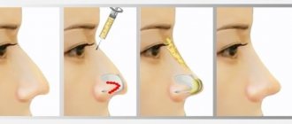
Milium
Milium, or whiteheads, is a retention cyst of the epidermis with a predominant localization on the face. It is a white, round, dense nodule with a diameter of 1-2 mm. Treatment. The only method of surgical treatment is to open the apex with a scalpel and remove the contents of the cyst. Healing without scar formation.
Keratoacanthoma
Keratoacanthoma is a rapidly growing dense hemispherical node, the size of which can exceed 5 cm. In the center of the tumor there is a crater-shaped depression filled with horny masses. Keratoacanthoma usually occurs on exposed areas of the body in people over 50 years of age. There are indications of a viral nature, the effects of prolonged insolation, trauma, exposure to tar and resins. After 2-3 months, the tumor resolves spontaneously, and a scar may appear in its place. Differential diagnosis is made with squamous cell skin cancer. Keratoacanthoma can be externally indistinguishable from squamous cell skin cancer, so this tumor is classified as both benign and facultative precancerous diseases.
Treatment. Usually resort to excision of the tumor. If the diagnosis is reliably established, electrocoagulation and cryodestruction with liquid nitrogen can be used instead of surgery.
Neurofibroma
Neurofibroma is a benign tumor originating from nerve endings, a soft pinkish-brownish tumor of various sizes, often pedunculated. Occurs on any part of the body except the soles and palms. Multiple neurofibromas are a sign of neurofibromatosis (Recklinghausen's disease). This disease is inherited in an autosomal dominant manner and occurs in adolescence. Over the years, the number and size of neurofibromas increase. Patients often complain of itching. If the tumor bothers the patient, is painful, impedes movement, or leads to disruption of certain functions, its excision is indicated.
Nevus of the sebaceous glands
Nevus of the sebaceous glands is a common developmental defect. It is usually an elongated, hairless neoplasm with a smooth surface, which becomes warty or nodular during puberty. Localized on the face or scalp. In approximately 15% of patients in adulthood, various benign tumors of the skin appendages and basal cell skin cancer develop from the nevus of the sebaceous glands. Differential diagnosis is carried out with linear epidermal nevus. Excision of the nevus is recommended in adolescence.
Fibrous nasal papule
Fibrous nasal papule, or involutive nevus, is a small dome-shaped elevation at the tip of the nose, almost indistinguishable in color from the surrounding skin. Occurs in older people. Differential diagnosis is carried out with basal cell skin cancer, neoplasms of skin appendages.
Treatment. For cosmetic purposes, as well as in case of difficulties in making a differential diagnosis, the tumor is cut off with a sharp scalpel at the skin level and sent for histological examination. The wound heals by secondary intention.