A burn is a common injury that can occur at home or at work. You can burn your skin with boiling liquid or steam, hot objects and particles of molten metal, aggressive caustic acids, alkalis and other chemicals. Electric shock and exposure to harmful ionizing radiation also cause damage to the skin. For small and shallow burns, conservative treatment is carried out. If the area of damage is extensive, and the damage affects deep skin structures, the only effective treatment method is skin transplantation after the burn.
Burn degrees
Based on the depth of tissue damage, burns are classified into 4 degrees.
- Damage to the surface layer (epidermis), accompanied by pain, redness and slight swelling of the burned area. Heals in a few days without special treatment.
- Damage to the epidermis and upper layer of the dermis, manifested by severe pain, swelling, redness, and the formation of blisters filled with clear serous fluid. With proper drug therapy, the injury heals in 10-14 days.
- A 3rd degree burn is classified into 2 types. Degree 3A is characterized by damage to the epidermis and dermis, the formation of large blisters with cloudy liquid inside, and a scab (crust) on the burned surface. Degree 3B is diagnosed when all skin layers are affected and subcutaneous fat is partially damaged. The resulting blisters are filled with bloody fluid.
- The most severe injury, accompanied by charring of the skin, muscle and bone tissue, and complete loss of sensitivity due to the destruction of nerve fibers.
Even a small but very deep burn is dangerous due to the penetration of infection and its spread through the bloodstream throughout the body. The results can be sepsis and death.
Indications for transplantation
Skin grafting surgery is called dermoplasty (otherwise known as skin grafting, transplantation), and is prescribed in situations where it is impossible to restore burned tissue by other means.
Mandatory indications for dermoplasty are:
- 3B and 4 degree burns with a affected area of more than 2.5 cm;
- 2-3 degree burns with a large area of damage;
- formation of rough scar tissue, severe skin defects;
- trophic ulcers on the burned area.
2nd and 3A degree burns are borderline and are usually treated conservatively. Less commonly, to speed up regeneration and prevent complications, the doctor may recommend skin grafting.
For deep burns that occupy a large area, the operation cannot be performed until young granulation connective tissue has formed on the wound surface, so transplantation is carried out no earlier than 20-30 days after the injury. For small wounds with smooth edges and no areas of necrosis, plastic surgery can be performed within the first week after injury.
In children
Particularly severe burn injuries to the skin are diagnosed in children - according to statistics, more than half of childhood victims underwent dermoplasty. Skin grafting after a burn in children is mandatory, otherwise:
- the formation of rough scars, constrictions, noticeable skin defects causing functional impairment, physical discomfort and psychological disorders;
- improper formation of the musculoskeletal system (due to uneven stretching of healthy and scar tissue, tendons and muscle fibers become twisted).
Features of care and rehabilitation after transplantation
The recovery period can be divided into 3 periods. The first is 2-3 days after the procedure, when the skin adjusts to itself. The second stage is regeneration, which takes 2-2.5 months. During this period, the area with the transplanted skin should be protected from various types of damage. The bandage is removed only with the consent of the doctor.
Wound care is an important part of postoperative care. The procedure is performed only in the clinic using sterile materials. For treatment at home, the doctor prescribes painkillers and also uses special ointments to maintain water balance in the wound. The main thing is not to allow the skin to dry out at the transplant site, otherwise severe itching will be felt. The recommendations given by the doctor before discharge are as follows:
- Timely change of dressing;
- bed rest;
- Do not wet the wound;
- Drinking;
- giving up alcohol;
- taking vitamins;
- Proper nutrition.
The third stage of recovery is rehabilitation. It takes 3 months for complete recovery. If you follow all the doctor’s instructions, the recovery period passes quickly and without visible complications. After this, the person will be able to return to their normal lifestyle.
Contraindications
Contraindications to dermoplasty are:
- absence of young connective (granulation) tissue with extensive damage;
- areas of necrosis on the wound;
- inflammation in the affected and nearby healthy areas;
- discharge of serous or purulent exudate;
- multiple hemorrhages, accumulation of liquid or coagulated blood in soft tissues (hematoma).
Relative contraindications include the unsatisfactory physical condition of the patient:
- burn shock;
- loss of a large volume of blood;
- exhaustion;
- low hemoglobin and poor indicators of other laboratory tests.
Preparation for dermoplasty
Primary dermoplasty is performed 3-4 days after the burn with a small wound area and does not require preparatory measures.
But more often, secondary (20-30 days after injury) skin grafting is performed after a burn. In this case, preparation of the wound for surgical intervention is mandatory: mechanical cleansing and drug therapy aimed at removing purulent and necrotic contents, preventing or treating infectious complications that have already arisen. Measures are also taken to stabilize and improve the patient’s general physical condition.
The preparation stage includes:
- prescription of vitamins and restoratives (to increase the body's resistance);
- systemic antibacterial therapy;
- the use of local antiseptics and antibacterial drugs (the application of ointments under the bandage is stopped 3-4 days before the planned date of transplantation, since remaining particles of drugs may complicate the engraftment of the graft);
- antiseptic baths with a solution of potassium permanganate or furatsilin;
- UV irradiation of the wound surface;
- blood transfusion (blood transfusion) or plasma transfusion (carried out according to indications).
Local treatment of burn victims on an outpatient basis
Burns are one of the most common types of injuries. In Russia, more than 400 thousand patients with thermal burns are registered annually. Moreover, only 30% of them require hospitalization; the remaining patients are treated in the clinic. In addition, most patients with the consequences of thermal injury after discharge from the hospital also continue treatment and rehabilitation on an outpatient basis. Therefore, effective treatment of burn victims depends not only on the combustiologist working in the burn center. In many ways, qualified medical care for burns at the outpatient stage determines their further course, the possibility of developing complications and the outcome of the injury.
Local treatment of burns is carried out at all stages of evacuation and treatment of burned victims in accordance with the established volume of medical care for each stage.
First aid to burn victims should be provided immediately, already at the scene of the incident, and begin with stopping the action of the thermal agent and, if possible, removing all materials in contact with the burnt surface (clothing, jewelry, etc.). Further, for local burns up to 10% of the body surface, it is necessary to cool the damaged areas of the skin for at least 15-20 minutes with water or the use of cold objects. Immediate, no later than 30 minutes after injury, cooling the burned surface reduces the time of tissue overheating, preventing the action of the thermal agent on deeper tissues. Cooling reduces swelling and relieves pain, and has a great influence on the further healing of burn wounds, preventing deepening of the damage. The patient should also be given painkillers and antihistamines, in case of extensive burns, warmed up and, if there is no vomiting, given something to drink. During the period of transportation of victims to a medical facility in the presence of limited burns, the primary dressing may be a dry aseptic dressing; for extensive burns, standard contour dressings or sterile sheets are used for these purposes. The primary dressing should not contain fats and oils due to subsequent difficulties in cleaning wounds, as well as dyes, because they can make it difficult to recognize the depth of the lesion.
Ready-made first aid dressings can be used to bandage burnt victims. Among modern first aid dressings and subsequent treatment, especially for patients with limited burns of II-IIIAB degrees, modern wound coverings of the Activtex and APPOLO series, as well as aerosol preparations (Amprovisol, Olazol, Panthenol), can be used.
In clinics and outpatient clinics, medical care can be provided to patients with thermal injuries, including for urgent indications and in order to prepare the victim for transportation to a specialized or surgical hospital. In this case, treatment tactics are determined by the possibility of continuing it on an outpatient basis or the need for hospitalization in a hospital. The criteria for hospitalization of burnt victims are the extent and depth of burns, their localization, the presence of thermal inhalation injury, concomitant injury and concomitant diseases, as well as the age of the victim. It should be noted that during the initial examination it can be difficult to determine the depth of the burns. Most often, the true depth of the burn can only be determined after 5-7 days.
For an outpatient surgeon, it is necessary to have a clear understanding of the indications for hospitalization of burn victims:
Deep burns of IIIB-IV degree. Superficial burns of I-II degree - more than 15% of the body surface. Borderline burns of IIIA degree - more than 5% of the body surface. Burns of special localizations (face, hands, feet or genitals). Thermal inhalation damage. Burn shock .General electrical trauma. Combined or concomitant trauma. Wound infectious complications.
It is known that burn wounds are always infected. The development of inflammation in IIIAB-IV degree burns is a stage of the wound process and is caused not so much by the influence of microflora, which is always present in a burn wound, but by the natural processes of limitation and rejection of dead tissue. Therefore, the often used term “infected burn” is inappropriate. The concept of a wound infectious complication of burns includes conditions when the purulent process spreads beyond the primary lesion, leading to the development of both local (abscess, phlegmon, thrombophlebitis, etc.) and general (pneumonia, sepsis) infectious complications.
Indications for outpatient treatment of burnt patients
Adult patients with superficial I-II degree burns can be treated on an outpatient basis if the affected area does not exceed 10-15% of the body surface, and for borderline IIIA degree burns - 5% of the body surface, outpatient treatment of small-area deep pinpoint burns is also possible, for example from splashing hot oil. For this category of patients, the main thing is local conservative treatment of burn wounds.
Goals of local treatment of burns I-II-IIIA degrees
Local treatment for burns of I-II-IIIA degrees should be aimed at creating the most favorable conditions for their healing in the optimal time and provide protection of the wound from mechanical damage and infection, and, if necessary, effective treatment of wound infection and stimulation of reparative processes.
Primary toilet of a burn wound
Local treatment begins with the primary toilet of the burn wound. After anesthesia, carefully, in a minimally traumatic way, clean the burn surface from contamination, foreign bodies and scraps of epidermis. The wound is treated with antiseptic solutions. Opened bubbles are removed. Large unopened blisters are cut at the base and, without removing the epidermis, the contents are evacuated. Smaller bubbles do not need to be opened. In case of suppuration of the contents of the blisters, the exfoliated epidermis should be removed. It is better not to remove coagulated dry fibrin, since this will injure the underlying tissues; treatment in these cases is carried out under a thin scab. Tetanus prophylaxis is also recommended for all burn victims.
Local treatment of superficial and borderline burns
Treatment of burns of I-II-IIIA degrees can be carried out using both open and closed methods. Moreover, depending on the nature of the created wound environment, these methods are implemented using dry or wet methods. Treatment of burn wounds with the open method is possible on the face, in the genital area and perineum, where bandages make care and physiological functions difficult. In these cases, iodopirone solution, aerosols, and also creams containing silver preparations are used. However, in an outpatient setting, the method of choice is the closed method using various dressings.
For victims with local I-II degree burns, it is possible to use the aerosol drug Acerbin, which has an antiseptic and wound-healing effect. It can already be used when providing first aid. Acerbine, if necessary, is applied several times a day to the wound, which is covered with a sterile bandage soaked in the solution.
After removing the exfoliated epidermis in the absence of signs of infection, it is sufficient to use atraumatic dressings (Parapran, Voskopran, Branolind and others), a single application of which to superficial burn wounds is sufficient for subsequent epithelization under the bandage. You can use other wound dressings (Activtex, Appolo), as well as dressings with antiseptic or antibiotic solutions, water-soluble ointments or creams based on silver sulfadiazine.
For burns of IIIA degree, as well as small deep burns of IIIB degree, treatment can begin with wet-drying dressings with solutions of Iodopirone (Betadine, Povidone-iodine), which promote the formation of a thin scab consisting of necrotic layers of skin and fibrin. The disadvantages of the latter include pain during the first dressings. Under a dry scab, burns can heal without suppuration. In cases where a dry scab cannot be formed, already a week after the injury its suppuration and rejection develop. Treatment should be continued with bandages with water-soluble ointments, and the sloughed scab should be removed during dressings.
At the same time, the use of gauze dressings as a carrier for drugs has a number of disadvantages. Removing dried gauze dressings on dressings, even despite soaking, leads to traumatization of the young epithelium and disruption of spontaneous epithelization of the wound, and the dressings themselves are painful. That's why
The use of atraumatic “mesh” coverings in outpatient settings helps solve the problem of epithelial trauma, reduces the pain of dressings and has a beneficial effect on the course of the wound process.
One of the types of modern dressings is Activtex wound coverings, which are a textile base impregnated with a gel-forming polymer and containing various medicinal substances in the form of a depot system. Depending on the drugs included in the composition, various types of coatings with antibacterial, analgesic and hemostatic effects can be used. The therapeutic effect of the components that make up the Activetex coatings manifests itself only in a wet state, and therefore it is necessary to moisten the dressing before use and then periodically 1-2 times a day. The advantages of Activtex anti-burn dressings are the absence of the need for their preliminary (immediately before use) preparation and prolonged healing properties.
Dressings should be performed at least 2 times a week. If the patient's temperature rises, pain and swelling in the wound area intensify, the bandage becomes wet with pus, it should be replaced more often. Dressings should be performed sparingly, without injuring the thin layer of growing epithelium, especially in the treatment of IIIA degree burns, when epithelization occurs from preserved skin derivatives.
A modern alternative to the dry method of local treatment, when epithelization of superficial burns occurs under the scab, is to create a moist environment on the wound. A humid environment has a beneficial effect on regeneration processes, while the dressings themselves are atraumatic.
A wet dressing method for local treatment of burns is implemented using various film, hydrogel, hydrocolloid and sponge dressings, as well as creams based on silver sulfadiazine.
In the treatment of superficial and borderline burns, various film dressings are highly effective. At the same time, the lack of drainage properties of film dressings requires more frequent (daily) dressings, especially with heavy wound discharge. Therefore, the use of occlusive dressings is contraindicated in the treatment of purulent-necrotic and infected burn wounds with microbial contamination of more than 104 microbial bodies/cm2.
In the treatment of limited superficial and borderline burns, dressings containing hydrogels are widely used. Form-stable hydrogel coatings (Gelepran, Hydrosorb) show good effectiveness in the treatment of burn wounds of II-IIIAB degrees, as well as long-term wounds. Apollo's mesh-based dressing with an amorphous hydrogel differs favorably from its Western counterparts by the presence of anilocaine as an anesthetic and iodovidone as an antiseptic. Hydrocolloid dressings (Hydrocoll) can also be used effectively.
The use of biological materials in the treatment of superficial burns of the first and second degrees is inappropriate. When treating borderline IIIA degree burns in a clinic, various sponge coatings (Algimaf, Algikol, Syuspur-derm, etc.) can be used. They do not require frequent dressings, absorb wound detritus and promote regeneration.
The use of hydrogel, hydrocolloid and sponge dressings due to the active sorption of discharge allows dressings to be performed after 2-3 days, and in the absence of discharge and the dressings are tightly fixed to the wound, they can be left until complete epithelialization and independent separation from the healed surface.
Creams with silver, produced under various names (Argosulfan, Dermazin, Ebermin), which maintain a moist wound environment, create favorable conditions for epithelization and are effective means for local treatment of superficial and border burn wounds, especially in combination with film coatings.
The use of these drugs is advisable for clinical signs of infection and severe suppuration of burn wounds. In addition to them, it is possible to use a solution of Iodopirone (Betadine) and Lavasept. In the treatment of infected burn wounds, gauze dressings with multicomponent ointments on a water-soluble (polyethylene glycol) basis (Levomikol, Levosin, Dioxidin, Iodopyron, Betadine), which can be used in phases I and II of the wound process, have proven themselves well.
With an uncomplicated course of the wound process, the correct choice of methods and means of local treatment, II degree burns heal 1-2 weeks after injury, IIIA degree burns - by 18-21 days. Long-term conservative treatment of non-healing burn wounds in a clinic is a mistake. All burn wounds that have not healed within 3-4 weeks, especially those occupying an area of more than 0.5% of the body area, are deep and most likely require autodermoplasty.
Treatment of burnt patients discharged from the hospital with small residual wounds remaining after superficial burns and in areas of surviving grafts in those operated on for deep burns is carried out according to the general principles of local treatment indicated above.
The criteria for successful treatment of burnt patients is not only wound healing, but also good functional and cosmetic results. According to various studies, in 10% of cases after II degree burns, in 55-62% after IIIA degree burns and in 30-40% of cases after autodermoplasty for deep IIIB-IV degree burns, hypertrophic and keloid scars develop.
It should be noted that the main task in the fight against pathological scars is not so much their treatment as the prevention of scar formation. Carrying out anti-scar measures is most effective for “fresh” scars during the period of their formation. Most conservative treatment of burn scars occurs on an outpatient basis. Typically, a comprehensive course of treatment is carried out over 6-12 months, combining various methods: general and local drug treatment, wearing compression clothing, physiotherapy, massage, physical therapy, balneological treatment, etc.
When carrying out rehabilitation of burn survivors, it is important to take into account that many patients have mental disorders associated with the consciousness of their inferiority. This circumstance requires a special approach and often the need for drug correction. In addition to the development of scars and deformities, patients who have suffered a severe burn disease may have various dysfunctions of internal organs, requiring the involvement of appropriate specialists in the examination and treatment of such victims. Our country does not yet have a unified system for the rehabilitation of burn victims. Training clinic surgeons in the specifics of treatment and rehabilitation of burn victims with the involvement of specialists from burn departments may be one of the ways to solve this problem. More attention should also be paid to improving the system of medical and labor examination of burn survivors and their employment.
After 6-12 months of outpatient conservative treatment after the scars have “matured,” the patient, if necessary, undergoes reconstructive surgery. In cases of rapid progression of microstomia and eyelid eversion, the patient is advised to undergo reconstructive surgery urgently.
The emergence and introduction into clinical practice of new drugs and wound dressings makes it possible to improve the quality of medical care for patients with thermal injuries, including in a clinic, subject to compliance with the appropriate protocols and standards of treatment.
Operation technique
OLYMPUS DIGITAL CAMERA
Skin grafting for burns is carried out in several stages.
- A special instrument is used to collect material for transplantation. This stage is omitted if the graft material is not your own skin.
- The burn wound is prepared for transplantation: the wound surface is cleaned, necrotic tissue is removed, rough scars along the edges of the wound are excised, the wound bed is leveled, treated with an antiseptic, and irrigated with an antibiotic solution.
- The graft is aligned with the edges of the wound surface and, if necessary, fixed with catgut (self-absorbable suture material), a mixture of fibrin and penicillin solutions.
- The donor material is irrigated with glucocorticosteroid solutions to reduce the risk of rejection.
- Moist cotton balls and swabs are applied to the transplanted material, and a pressure bandage is placed on top.
Materials used
There are various synthetic and natural materials used for dermoplasty:
- autoskin – own skin taken from an unburned area of the body that is usually hidden by clothing (inner thigh, back, buttocks);
- allo-skin – cadaveric skin (taken from a dead person), preserved for subsequent use in transplant surgery;
- xenoskin – animal skin (usually pig skin);
- amnion – the embryonic membrane of the embryo of humans and higher vertebrates (reptiles, birds, mammals);
- artificially grown collagen and epidermal materials.
Skin grafting is most often performed by specialists using biological materials – autoskin and allo-skin. Amnion, xenoskin, and other synthetic and natural materials generally cover the wound surface temporarily in order to prevent infection.
When choosing the appropriate material, the depth of the burn is also taken into account. For grade 3B and grade 4 injuries, it is recommended to use your own skin, while for grade 3A injuries, plastic surgery is performed using allo-skin.
When transplanting your own skin, flaps taken from a certain area of the body can be used, which are not connected in any way to other tissues and organs. This type of surgery is called free plastic surgery. Surgery using the skin around the wound, which is moved and stretched over the burn area using microscopic incisions, is called non-free plastic surgery.
The quality and physical characteristics of the graft depend on the thickness of the flap. According to this parameter, donor materials are classified into 4 types.
- Thin flap (no more than 20-30 microns). Includes the epidermis and the basal layer of the dermis. It is characterized by poor elasticity, often wrinkles, and is easily damaged, therefore it is used very rarely for burns, mainly for temporary covering of the burn area.
- A flap of medium thickness, otherwise intermediate, split (from 30 to 75 microns). Consists of epidermis and dermis (full or partial layer). It is characterized by high strength and elasticity, practically no different from your own healthy skin. It is most often used for transplantation after burns; it is suitable for restoring skin on moving areas (articular surfaces), since it has sufficient elasticity and does not impede movement.
- Thick flap (50-120 microns). Includes all layers of the skin. Used for very deep wounds or damage to open areas of the body (face, neck, décolleté). It is transplanted only to areas where there is a sufficient number of capillaries that will connect to the vessels of the donor flap.
- Composite flap. In addition to the skin itself, it includes subcutaneous fatty tissue and cartilage tissue. Used for facial burns.
About the recipient site
After surgery, the recipient site may be covered with a pressure or vacuum bandage.
Pressure bandage
A pressure bandage will help the recipient site heal properly. It exerts pressure on the recipient area, due to which liquid does not accumulate under it. This allows the skin flap to adhere to the skin. It can be secured with silk sutures, a splint, a plaster cast or a support bandage. This will prevent the flap from moving.
Your surgeon or nurse will remove the pressure bandage approximately 5 to 7 days after surgery. Once the pressure dressing is removed, the recipient site will be covered with a Xeroform dressing.
If you have a cast, your surgeon will cut out a portion of it above the recipient site. This will allow him to examine the flap. The cast will be removed 10 days after surgery unless you have had other surgeries. If you have had other surgeries, you may need to wear the cast longer. An Ace® bandage, gauze bandage, or tape will be used to secure the Xeroform cast after the cast is removed.
You may be told to change the Xeroform dressing and gauze once a day until the flap is completely healed. Your nurse will teach you and your caregiver how to change the dressing.
Vacuum wound dressing
Instead of a pressure bandage, the surgeon may use a vacuum bandage on the recipient site. A vacuum dressing is a special dressing that suctions fluid from a wound, which speeds up healing.
The vacuum wound dressing will be removed 5 to 7 days after surgery. The surgeon will then examine the flap to ensure it has completely healed. If the flap has not healed completely, you may need to wear the vacuum dressing longer.
to come back to the beginning
Complications
One of the main complications of skin grafting is graft rejection, which can occur even after transplanting your own skin. The reason for this phenomenon is most often the presence of remains of pus, necrotic cells, and medicinal substances in the wound.
When rejected, complete or partial necrosis of the transplanted skin occurs. In this case, the dead tissue is removed, after which a re-transplantation is performed. If the rejection is partial, only the foci of necrosis are removed, leaving the engrafted tissue.
Other common complications of skin grafting include:
- bleeding of postoperative sutures;
- tissue compaction and scar formation along the edge of the wound surface (at the junction of healthy and donor skin);
- secondary infection of the wound (if the rules of preoperative preparation are violated or asepsis is not observed during surgery);
- sepsis;
- cracking of the donor skin, the appearance of ulcers and erosions on its surface, limitation of movements due to tightening, especially on the articular surfaces (occurs when the wrong material is chosen or the transplantation is not carried out in a timely manner);
- decreased sensitivity, atrophy (decrease in volume) of tissue in the transplant area.
Pros and cons of skin plastic surgery
Skin transplantation after a burn, like all other types of plastic surgery, has its advantages and disadvantages. The first include:
- protection of the wound from infection and mechanical damage;
- preventing moisture evaporation and loss of nutritional compounds through the open surface of the wound;
- more aesthetic appearance after healing of the wound surface.
The disadvantages of surgical intervention include:
- the possibility of rejection of the transplanted material (when transplanting your own skin, the risks are much lower);
- the likelihood of developing other complications;
- psychological discomfort of the patient (for example, if the donor material is cadaveric skin or tissue taken from an amputated limb).
Skin transplantation for burns. Advantages and disadvantages of the method
The main advantages include:
- formation of a protective layer against infections and mechanical damage;
- protection from increased moisture and loss of essential nutrients through the open wound area;
- aesthetic appearance.
Disadvantages of the method:
- risk of donor site rejection. The risk of rejection is significantly reduced when using your own tissue;
- risk of other complications.
Price
The cost of skin grafting is determined by various factors: the area of the damaged surface and the complexity of the specific operation, the type of materials and anesthesia used for transplantation, other pharmacological agents, the qualifications and professionalism of the surgeon, the location and reputation of the clinic. Depending on these factors, the price of the operation can range from 10,000 to 200,000 rubles.
Skin plastic surgery after a burn is not a complex or lengthy operation. But in some situations, graft engraftment after transplantation proceeds poorly, even if the donor material is one’s own skin. Detailed preparation for plastic surgery, correct calculation of the timing of its implementation, and appropriate care during the rehabilitation period are mandatory conditions for successful skin plastic surgery and help minimize the likelihood of developing postoperative complications.
Classification of grafts for transplantation
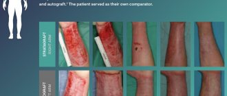
For skin transplantation, it is preferable to take material from the patient himself (autograft). If this is not possible, a living or deceased donor (allograft) is used. Sometimes doctors use the skin of animals, especially pigs. Developed clinics practice growing synthetic skin - an explant.
Depending on the depth of the lesion, the material for transplantation is divided into three types:
- thin - up to 3 mm. The biomaterial consists of the upper and additional layers of skin and has a small amount of elastic fibers;
- average - 3-7 mm. It consists of a mesh layer rich in elastic fibers;
- thickness - up to 1.1 cm. It covers all layers of the dermis.
The choice of material depends on the location of the burn, its size and the individual characteristics of the body.

