Keratoma is a collective name for similar benign skin tumors that have the same nature of origin. All keratomas are formed from superficial cells of the epidermis - keratinocytes and are characterized by the growth of the outer layer of the epidermis with excessive keratinization. Visually they look like single or multiple rough plaques, nodes or spots of different colors. They can be localized almost throughout the body, except for the skin of the palms and soles. Not contagious - not transmitted from person to person.
Keratomas most often occur only in adults. The main peak of their growth occurs at the age of 50+ and older. Slightly less often, formations are found in 30-year-old patients and very rarely in twenty-year-old patients.
Types of keratomas
There are several types of keratomas. Some of them are benign and may disappear spontaneously. Others may become malignant.
- Senile or senile keratoma is the most common type of neoplasm. In the initial stages, it looks like brown spots, which are most often located on open areas of the skin. As the spot grows, it becomes convex and loose, peeling and possibly even weeping occurs on its surface. Senile keratoma rarely disappears on its own, but can transform into a precancerous neoplasm - a cutaneous horn.
- Seborrheic keratoma. This neoplasm grows slowly, it all begins with the formation of spots from light beige to brown, which over time become covered with scales. These spots increase in size and may merge with each other. As they grow, the crusts begin to thicken, which subsequently peel off and crack. Seborrheic keratoma does not malignize, but does not disappear on its own.
- Horny keratoma, also known as cutaneous horn. A dense formation that can reach a height of several centimeters. An inflammatory rim is visible on the skin along its perimeter. Prone to malignant transformation.
- Follicular keratoma. It is more common in women and is located around the hair follicles. It looks like a convex light pink or light gray node, surrounded by a pink ring with a slightly rough surface. Its size can reach one and a half centimeters. In the center of the neoplasm there is a funnel-shaped depression. More often this is a single formation, which is mainly localized in the scalp, or on the cheek, or near the red border of the lips. Follicular keratoma grows slowly and does not become malignant. But it can quickly recur after removal.
- Actinic or solar keratoma. The provoking factor for its appearance is ultraviolet radiation - regular, long-term insolation. More often, these are multiple formations that are localized mainly in open areas of the body: face, forearms, shoulders, etc. The process begins with the appearance of small pink papules, on the surface of which flaky crusts are visible. They quickly transform into plaques similar to moles, along the periphery of which an inflammatory skin reaction is visible. The scales become dense and rough, but are easily removed. Solar keratoma is one of the most frequently degenerating skin tumors, and therefore belongs to precancerous skin diseases. Even if it resolves on its own, there is a possibility of its recurrence in the same place.
- Angiokeratomas. In appearance they resemble hemangiomas. These are nodular dark red or bluish neoplasms with blurred boundaries. Due to keratization, their surface peels off. No spontaneous resolution or malignancy is observed. May occur in childhood and adolescence.
Why you may need to remove a keratoma
Keratoma is a benign neoplasm on the skin, which consists of epidermal cells. It can be either small in size (a few millimeters) or quite large (up to 5 centimeters in diameter). Keratomas are usually slightly darker than the natural color of the skin or have a brown color. Over time, the surface of the keratoma becomes rough and rough.
The neoplasm is formed from human skin cells, which gradually layer on top of each other. At the same time, the upper layer becomes horny, skin scales die and fall off under mechanical stress, for example, during washing, but the deeper layers of the dermis also become keratinized. The process of continuous cell renewal leads to the fact that the keratoma does not disappear on its own, but can cause inconvenience for a long time.
Under normal conditions, the number of dead and new cells on human skin is balanced in such a way that the epidermis has time to renew itself in time and maintain a healthy state. If this balance is disturbed and the cells become keratinized at a faster rate than they are renewed, then various growths appear on the skin, including benign ones, such as keratomas.
Benign neoplasms on the skin are not dangerous to human health, since they consist of normal, unchanged cells. However, there is a fairly high probability that these formations will degenerate into malignant ones when exposed to a number of negative factors. These include, for example, excessive sun exposure or contact with toxic chemicals.
Keratomas can occur on any area of the skin. They can be single or form groups. Most often, keratomas are located on the neck, shoulders and arms. Less common on the lower body.
Currently, the reasons for the appearance of these formations on the skin are not fully understood. Nevertheless, scientists have proven that prolonged exposure to the sun and visiting a solarium promotes the growth of keratomas.
How to remove keratomas
In our clinic, keratomas can be removed on the day of treatment, after dermatoscopy and determination of the nature and benignity of the formation. Our doctors practice several modern methods of removing keratomas. The choice of removal method is made based on the location of the tumor, its size and the risk of malignancy.
Why is keratoma dangerous and why is it necessary to remove it?
Keratoma itself does not pose a threat to human life and health. However, due to the fact that over time it is increasingly exposed to negative factors, such as mechanical damage and exposure to ultraviolet radiation, this benign neoplasm can degenerate into a malignant tumor.
Sometimes a keratoma can cause significant discomfort to a person. Itching, burning sensation, inflammation, bleeding, pain - all these are alarming signs. The appearance of these symptoms may indicate that the process of degeneration of the keratoma into a malignant neoplasm has begun.
If you notice new moles, spots on your skin, or strange growths, be sure to visit a dermatologist-oncologist who can conduct research and determine whether the formations on the skin are malignant. Next, it is important to undergo regular medical examinations to prevent the occurrence of cancerous tumors.
Also, in a medical institution, you may be offered to remove keratomas on the face and other parts of the body and get rid of the need to regularly see a doctor.
Removal of keratomas with laser
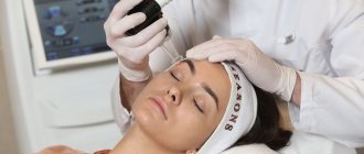
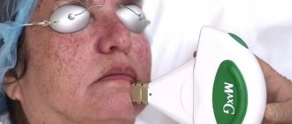
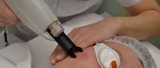
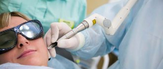
To remove large keratomas and keratomas localized in the face and dorsum of the hand, our doctors use the Fotona SP Dynamis erbium laser. The technology of laser removal of keratomas is based on heating and destruction of pathological tissues under the influence of laser radiation. As a result, necrosis of the neoplasm occurs with the formation of a dry scab. Underneath it, epithelization of the wound occurs, and after the crust falls off, a delicate skin remains in this place, which then transforms into normal epithelium.
Possible complications and consequences of laser intervention
Laser surgery of neoplasms can leave scars - small and unnoticeable, or larger ones if the keratoma was initially large. In addition, patients note the appearance of numbness in the tissue at the site where the tumor was removed.
Such manifestations are natural consequences of the procedure.
It is necessary to pay attention to some dangerous symptoms of poorly carried out destruction of the tumor, for example, the appearance of redness around the crust formed after removal, severe itching or rash at the site of the keratoma.
If the crust has fallen off on its own, and underneath it an ulcer in the form of a red spot with an uneven surface is found that has not healed for a long time, you need to visit a dermatologist as soon as possible.
In addition, allergic reactions or dermatitis may develop around the procedure site due to constant wearing of a bandage, as well as the use of various ointments on the skin. At the first manifestations of dermatitis or allergic rashes, you must stop treating the area with any medications and also consult a dermatologist.
Removal using radio waves
Radioknife, or radio wave surgery (Surgitron device) is the most universal method for removing dermatological tumors, which can be used even in hard-to-reach locations. As in the previous case, the method is based on heating and evaporation of cells under the influence of radio frequency waves. The technology allows for targeted effects down to several millimeters. The surrounding tissues are not affected. Another advantage of the method is a good cosmetic result - healing occurs practically without the formation of noticeable marks and scars.
Wound surface care techniques
After laser destruction of the keratoma, a characteristic dark brown crust forms in its place. It covers the surface of the wound, and underneath it the tissue healing process is actively occurring. It is strictly forbidden to rip off or comb the scab, as this opens the door for pathogenic microorganisms to enter the wound until the need for it disappears - then it disappears on its own. For the first two days, it is not recommended to wet the wound site.
Postoperative care includes the following rules: once or twice a day, the procedure site must be washed with water and baby soap, after which it is necessary to treat the surface with antiseptic drugs and healing ointments. Next, a sterile gauze bandage is applied to the wound. If possible, it is better not to use an adhesive plaster, as it restricts air flow into the wound, thereby slowing down its healing.
Complete skin restoration occurs after 4-6 weeks. During this entire time, it is prohibited to visit the bathhouse, sauna, or swim in pools or open reservoirs. Exposure to ultraviolet rays is also undesirable, and not only during postoperative recovery, but also in the next 6-8 months, in order to avoid recurrence of keratoma, so it is better to forget about beaches and solariums for a while, and go outside in clothes or a hat that covers the affected area and treat it with sunscreen with a high UV filter.
Removal of keratomas using diathermocoagulation method
In this case, the removal of pathological tissues is carried out under the influence of very high temperatures (about 2500 oC) using the Plasmaskin apparatus. The advantages of the method include non-contact (which eliminates the risk of contamination by infections) and a disinfecting effect.
After removal of the keratoma, observation by a dermatologist is necessary.
IMPORTANT!!!Given the frequent cases of degeneration of keratomas, especially multiple ones, we advise you to keep these neoplasms under the supervision of doctors!!!
Come to our clinic. We will conduct a diagnostic study of the keratoma, select the optimal destruction method for you, and professionally carry out the removal procedure.
Timely treatment
The best way to fight cancer, one of the most terrible diseases facing humanity, is prevention. As soon as a keratoma appears, it is advisable to immediately, without delay, consult a dermatologist for laser removal. The smaller and more insignificant the skin tumor, the greater the likelihood that after its removal there will be no trace left on the body. Well, of course, such a step will significantly reduce the risk of malignant degeneration.
At the moment, only one way to treat keratoma is known - it must be removed.
This is a radical method, which can only be carried out efficiently by a qualified specialist. If suspicious growths are detected on the skin, you should contact specialized clinics and centers such as SkinLazerMed. The best option is to consult a dermatologist or dermato-oncologist.
Attempts to get rid of keratomas on your own can be fraught with the formation of unsightly scars at best, and the development of skin cancer at worst.
Preparation
It is important to undergo an examination by a dermatologist. First, the doctor will make an accurate diagnosis. Secondly, he will recommend a suitable removal method. Despite the versatility of the laser, some cases require the use of other methods. In addition, there are contraindications to any interventions, so it is important to consult a doctor and talk about your health status, and in many cases, undergo diagnostics. Otherwise, no specific preparation is required. It is best for women to schedule the intervention after menstruation, as sensitivity may increase during menstruation.
Post-procedure care
In order for the result of removing a mole, wart or any other formation to be predictable and recovery to be quick, you should adhere to the following rules:
- Do not wet the treatment area with water for 2-3 days until a thick crust appears.
- Do not be exposed to ultraviolet rays - artificial (solarium) and natural (sunlight) for 2-3 weeks after the procedure. Next, it is advisable to always protect the area with a cream with an SPF of at least 35.
- If the formation has been removed from the face, you should avoid foundations and powders for 3 weeks.
- Refusal to visit saunas, swimming pools and baths to minimize the risk of infection of the treated area for 1 month.
After the procedure itself, the specialist will tell you what antiseptic agents can be used to clean the skin area and how soon the crust should fall off (usually up to 7 days). After it has disappeared, you will need to apply restorative products to speed up the healing of the skin.
The use of adhesive plaster over the formed crust is contraindicated, as is the use of fat-based ointments or thick creams - the “greenhouse effect” will only slow down regeneration.
Service cost
Price information is provided in our price list. The full cost of the keratoma removal service depends on the method used, as well as the size and number of tumors.
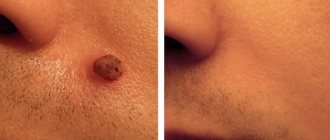
We suggest you evaluate in the photo the results of treatment of skin formations of this type at the Podology Clinic. The use of modern technological methods, as well as the efforts of our highly qualified doctors, help to perform the operation quickly and without the risk of noticeable scars.
If you need to get rid of keratoma, contact the Podology Clinic. You can make an appointment by calling +7 (495) 640-68-10.
Causes of skin keratinization
This disease is not congenital and occurs today in young people, starting from 30-35 years old. Single or multiple keratomas most often appear on the skin after 40-50 years. The main reasons for their formation are:
- age-related disruption of the mechanism of cell division and skin pigment production;
- abuse of sunbathing;
- malfunctions of the immune system;
- hormonal disorders associated with menopause;
- endocrine pathologies
- hereditary predisposition.
Prices for keratoma removal in Moscow are quite affordable, so if you notice such a tumor, sign up for a consultation with a specialist at the Podology Clinic. A surgeon or dermatologist will diagnose and remove the formation.
Surgical excision of keratoma
Surgical removal of keratomas is performed under local anesthesia using a scalpel. A similar procedure is prescribed in the following situations:
- Impossibility of carrying out other destructive methods due to certain reasons;
- Malignancy of the neoplasm;
- The presence of large, extensive growths;
- Lack of positive results after using other methods of combating keratomas.
The disadvantages of surgical removal of skin lesions include:
- Long and painful recovery period;
- Extensive damage to surrounding healthy tissue;
- High risk of infection and bleeding of the postoperative wound;
- The need for sutures;
- Formation of scars and scars.
To facilitate the rehabilitation period and reduce the risk of postoperative complications, the patient may be prescribed antibacterial and painkillers.
