According to experts, keratoma is not a hereditary pathology, but appears under the influence of certain conditions. The most common cause of the formation of growths is considered to be prolonged exposure to the sun. As a result of the influence of excessive amounts of ultraviolet radiation on the epidermis, cells begin to become keratinized, thereby forming a keratoma. Then it becomes harder and changes color.
Keratoma does not pose a threat to human life and health, so you can live with it. However, there is a possibility of the growth degenerating into a malignant tumor. In addition, a keratoma can cause a person certain aesthetic and psychological discomfort. Due to these characteristics of the neoplasm, there is only one conclusion - the keratoma should be removed. This will allow a person to get rid of complexes and prevent the development of cancer. Keratoma removal should be carried out in a clinical setting by a specialist and only in a way that is most suitable for the patient, taking into account his individual characteristics and indications.
Destructive methods for removing keratomas
Compared to medical methods of treating keratomas, surgical excision of skin tumors is considered the most effective way to combat growths. In modern medicine, there are several destructive methods for treating pathological formations. These include:
- Radio wave excision of growth;
- Removal of keratoma with liquid nitrogen;
- Cauterization of the growth with electric current (electrocoagulation);
- Laser burning of skin tumors;
- Surgical treatment of growths.
The most effective method for removing skin tumors is selected by the doctor after a preliminary examination and assessment of the clinical characteristics of the growth.
Traditional medicine in the treatment of keratoma
Traditional medicine recipes can be used as additional therapy and only after consultation with a doctor!
Celandine ointment and a mixture of fir and sea buckthorn oil will help slow down the growth of the tumor. You need to lubricate the keratoma three times a day for two weeks. In parallel with the main treatment, you need to adjust your diet - doctors recommend consuming more citrus fruits, blueberries, beans, soy and green tea. According to recent studies, these products contain large amounts of vitamin P, which slows down the development of keratoma.
With timely diagnosis and qualified treatment, the prognosis of the disease is favorable. For prevention purposes, doctors recommend minimizing exposure to direct sunlight and using body scrubs twice a week.
Radio wave removal of large keratomas
Eliminating growths on the skin using radio waves is one of the most precise and delicate methods of exposure. The procedure is carried out using a special radio wave surgery device - Surgitron. Under the influence of narrowly directed waves, intracellular fluid evaporates, followed by destruction of the formation. Thanks to the presence of a special applicator, the doctor can control the depth and intensity of the effect, which makes it possible to deal with keratomas of different locations and sizes. To relieve the patient from unpleasant and painful sensations, the procedure is performed using anesthesia.
The advantages of radio wave treatment of skin tumors include:
- Minimal damage to surrounding tissues;
- Efficiency and efficiency of the procedure;
- No postoperative complications or scars;
- Short rehabilitation period;
- Possibility of successful treatment of multiple and large growths.
What to remember:
- Keratoma is a benign neoplasm that appears due to mutation of skin cells and the growth of the keratinized layer of tissue.
- Ultraviolet light is the main provocateur of keratomas. Tanning with keratomas is contraindicated, both natural and in a solarium.
- For self-removal of keratomas, pharmacies sell special acid-based preparations.
- You can treat a keratoma yourself only after a medical diagnosis, making sure that it is not another tumor.
- Folk remedies are mainly aimed at eliminating discomfort from keratomas.
- Vinegar-based compositions remove growths. But they can lead to chemical burns.
Laser removal of keratomas
Cauterization of skin tumors with a laser is a fairly effective method of combating keratomas, which consists of exposing pathologically altered cells to laser beams, which leads to their destruction. Due to the coagulating properties of the laser, the risk of wound bleeding during or after the procedure is minimal. Exposure to high temperatures has a disinfecting effect, which avoids bacterial contamination and the spread of infection. During the procedure, healthy surrounding tissues are not damaged, which prevents the appearance of scars. Laser cauterization of growths is not recommended for patients with increased sensitivity to light exposure and tanned skin.
Causes of actinic keratosis
The main reason is hyperinsolation - excessive exposure to UV rays of a certain wavelength: from 280 to 320 nm, i.e. UVB. Moreover, the radiation is not one-time, but regularly affecting. Breakdowns and manifestations in the form of keratomas do not occur immediately. For decades, they do not appear and the skin retains its external structure. This is the latent period of the disease . With age, the skin begins to change and a pathological focus is formed - the manifested period .
Provoking factors contributing to the formation of keratosis and its development:
Climate impact. Living in a sunny region increases the risk of development tenfold. Most often, actinic keratosis affects residents of mountains, equatorial and tropical countries.
Work outdoors.
Age. The risk of developing senile keratoma increases after 50 years of age due to hormonal and immune changes.
Phototype. People with fair skin types get sick 3 times more often than people with dark skin. It has been noted that keratomas more often occur in people with freckles.
Sunburn. A powerful provocateur not only of skin hyperpigmentation, but also of more serious dermatological diseases with the risk of neoplasms.
Removal of keratomas with liquid nitrogen
Cauterization of the keratoma with nitrogen is one of the effective and common destructive methods of combating skin growths. The essence of removing a tumor with liquid nitrogen is to freeze the pathologically altered cells by using hardware or applying a cotton applicator soaked in liquid nitrogen to the problem areas. With the correct choice of intensity and depth of exposure, removal of large keratoma by cryodestruction allows achieving high aesthetic results. Incompletely removed growth roots can lead to recurrence of the tumor. Cauterization of small and single formations is carried out in one session. Treatment of multiple or large growths may require multiple procedures. After the initial exposure, the doctor evaluates the results of the operation and, if necessary, prescribes repeated cryotherapy.
The disadvantages of removing keratomas with nitrogen include the formation of a postoperative blister, which appears a few hours after cryodestruction and persists for 3-4 days. The patient should be careful and protect the bladder from damage and mechanical stress. Injuring it can disrupt the tissue regeneration process and lead to wound infection. It is also not recommended to pop a blister at home. If it causes significant discomfort and tightens the skin, you can consult a doctor who will open it and apply a sterile bandage.
Nitrogen removal is not recommended for people with allergic reactions to cold exposure, exacerbation of chronic diseases, an active form of herpes infection, acute inflammatory and infectious processes and cancer.
Rehabilitation period after keratoma removal
Removal of keratomas by any of the above methods involves a postoperative period, which lasts differently. The longest recovery process is observed after surgery. It can range from 2 weeks to 4 months.
After removing a keratoma using any of the destructive methods, it is important to adhere to the following recommendations:
- avoid contact of water and moisture on the affected area of the skin;
- do not remove the crust on the treated area of the dermis, as this can lead to the appearance of scars, scars and infection;
- try not to touch the inflamed epithelium and not expose it to ultraviolet radiation;
- use medications prescribed by a doctor;
- do not self-medicate, do not resort to traditional medicine unless your doctor approves it.
If you follow these recommendations, the regeneration process will be quick, painless and without complications.
After the keratoma removal procedure, it is important to come to an in-person appointment with a doctor for an examination 10–12 days later. The doctor will be able to evaluate the effectiveness of the applied technique and outline a plan for further action, as well as give recommendations for skin care.
In our clinic, you can remove keratomas using liquid nitrogen, as well as any other methods at an affordable price. After consultation, specialists will select the most suitable procedure and eliminate the skin defect.
Why clients choose Veronika Herba Beauty and Health Center:
- This is a beauty center where you can take care of yourself at a reasonable cost, while your face and/or body will be treated not by an ordinary cosmetologist, but by one of the best cosmetologists in Moscow. This is a completely different, higher level of service!
- You can receive qualified help at any time convenient for you. The beauty center is open from 9:00 to 21:00, seven days a week. The main thing is to agree with your doctor in advance on the date and time of your appointment.
Sign up for a consultation with a specialist by phone +7 (495) 085-15-13
, and you will see for yourself!
Electrocoagulation of keratome
Burning out skin growths using electrocoagulation is one of the destructive methods of combating keratomas, which is used in the treatment of fresh formations. For old keratomas, this method is ineffective. The essence of electrocoagulation is the effect of electric current on problem areas, which leads to destruction and necrosis of pathologically altered tissues. Since the electrocoagulation procedure can be quite painful, it is performed with the use of painkillers. When treating growths with electrocoagulation, the formation is completely eliminated without damaging healthy surrounding tissue. This makes the procedure quite popular in cases of suspected malignancy of the neoplasm.
Surgical excision of keratoma
Surgical removal of keratomas is performed under local anesthesia using a scalpel. A similar procedure is prescribed in the following situations:
- Impossibility of carrying out other destructive methods due to certain reasons;
- Malignancy of the neoplasm;
- The presence of large, extensive growths;
- Lack of positive results after using other methods of combating keratomas.
The disadvantages of surgical removal of skin lesions include:
- Long and painful recovery period;
- Extensive damage to surrounding healthy tissue;
- High risk of infection and bleeding of the postoperative wound;
- The need for sutures;
- Formation of scars and scars.
To facilitate the rehabilitation period and reduce the risk of postoperative complications, the patient may be prescribed antibacterial and painkillers.
Rehabilitation
Thanks to gentle laser removal, there is no need for a long recovery period. At the site of exposure, the skin becomes covered with a crust, under which healing proceeds.
The crust must not be scratched, wet, removed, or covered with cosmetics. The new skin that appears under the crust must be protected from exposure to sunlight. You need to purchase a high-quality cream with UV filters and apply it before going outside.
After complete healing, a healthy area appears at the site of the wound, which does not require special care. Wound infection and bleeding are completely excluded. This is due to the sterilizing effect of the laser on tissues, as well as blood vessels.
Preparation and performance of keratoma removal surgery
- Regardless of which method of combating growths is used, the patient undergoes a preliminary examination, which includes an examination by a dermatologist, a consultation with an anesthesiologist (provided that the growth is operated on using anesthesia), as well as a number of laboratory and instrumental studies. If a tumor is suspected of being malignant, consultation with an oncologist is necessary. During the diagnostic process, the doctor determines the presence of possible contraindications and allergic reactions, assesses the general health of the patient, as well as the location, size, prevalence and number of growths.
- Based on the information received, taking into account the patient’s wishes, the specialist selects the most suitable method for removing the skin lesion and sets a date for the operation. The procedure for excision of the growth does not require any special preparation. The patient comes to the clinic on the appointed day and, after consulting with the doctor, goes to the operating room.
- Before starting the procedure, the doctor treats the skin with antiseptic drugs, after which he uses an anesthetic gel, spray or injection (the dose and method of administration of the anesthetic is determined by the anesthesiologist at a preliminary consultation). Then the doctor, taking into account the characteristics of the growth (its scale, quantity, location) and depending on what destructive method is used, adjusts the devices to the necessary parameters and selects the appropriate devices. This will allow you to control the intensity and depth of the impact. After this, the doctor begins to remove the pathological growths using one of the above destructive methods. The removed growth is sent for histological examination to determine the nature of the neoplasm.
- At the end of the procedure, the surgical field is treated with an antiseptic. If necessary, stitches and a sterile bandage are applied. The patient is prescribed general strengthening, anti-inflammatory drugs, as well as healing and restorative agents for external use. This will speed up the healing of the epidermis, avoid bacterial infection and inflammation of the surgical wound.
Classification
Keratomas are classified as follows:
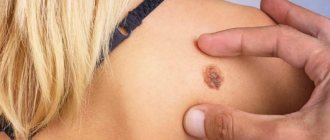
- Seborrheic keratoma appears most often on the chest, back and scalp. Initially, this is a yellow-brown spot, the diameter of which can reach 3 cm. Over time, the surface of the formation becomes dense, and scales appear on it. The color changes to dark brown or black. Externally, this skin pathology looks like dried raisins stuck in the skin.
- Senile keratoma affects the skin of people over 35 years of age. A spot with a diameter of up to 2.5 centimeters appears on the skin. Over time, it thickens, becomes crusty, cracks, and changes color from yellow to black. The growth grows slowly, but over time it can protrude above the skin by a centimeter or even more. Senile keratoma, as this type of neoplasm is also called, is often localized on the shoulders, head, and back. Less common on the face and neck. Senile keratomas can grow in colonies. They also often become inflamed due to physical damage.
- Horny keratoma, which dermatologists often call “cutaneous horn,” has an increased risk of degeneration. First, a gray or brown spot appears on the skin, which eventually becomes covered with scales. A convex tubercle with a rough surface is suitable for placement of the arms, chest, and back. But other parts of the body are no exception. Sometimes patients are diagnosed with multiple horny keratomas.
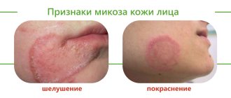
- Follicular keratoma appears on the cheeks, lips, nose, eyelids. The skin first turns pink, then small tubercles grow at the site where the spot appears.
- Angiokeratoma is a nodular formation of red, black or blue color. Its size can reach 10 centimeters, but most often the growth grows to 2-3 centimeters. Often such skin pathologies form on the back, abdomen, legs and genitals.
- At the first stage, solar keratoma looks like a flat mole. Gradually it begins to peel off. Scales form on its surface. Dense lesions are often found in men over 40 years of age. Location: back, face, feet. Education is rarely singular. As a rule, this is a whole colony of growths. In some cases, degeneration into squamous cell carcinoma is diagnosed.
The type of keratoma and its risks for the patient are diagnosed by a dermatologist after examining the formation.