Thanks to the work of the muscles, the eyelids close—blinking—which uniformly moistens the eye and removes foreign bodies that have gotten inside. Under the muscle tissue of the eyelids there is a dense fragment of collagen tissue - the cartilage of the eyelid, which maintains its shape and also ensures the strength of the eyelid structure. The cartilage includes the meibomian glands, which produce a specific fatty secretion that improves contact between the eye and the back of the eyelids and the closure of the eyelids. From the inside, the cartilage is tightly connected to the conjunctiva - a mucous membrane that produces mucin with tear fluid necessary to moisturize the eye and ensure the sliding of the eyelid folds along the eyeball. The eyelids have a rich blood supply network. The operation of the eyelids is controlled by the facial and oculomotor nerves.
Structure of the eyelids
The edges of the upper and lower eyelids form a palpebral fissure, in each corner of which the eyelids are connected to each other by certain ligaments. These ligaments firmly fix the cartilage of the eyelids directly to the walls of the orbit.
When closed, the edges of the eyelids fit tightly to each other. The edge of the eyelid consists of two ribs: anterior and posterior, in addition, it includes an intercostal space called intermarginal. The anterior edge of the eyelid is rounded and contains approximately 100 eyelashes, into whose bulbs the ducts of the sebaceous glands emerge, and between the eyelashes there are sweat glands. The intermarginal space, in turn, includes the excretory ducts of the meibomian glands. These glands produce a fatty secretion that provides lubrication of the edges of the eyelids, due to which the eyelids can close tightly and slide over the surface of the eye, and it also ensures the correct outflow of tears. In the inner corner of the intermarginal space of each eyelid there is a lacrimal papilla, the apex of which is the lacrimal punctum, through which tears should normally flow. At the posterior edge of the eyelid, the cut is sharp, which ensures tight contact with the surface of the eye.
The eyelid consists of two plates: the outer musculocutaneous one, and the inner one, which includes cartilage and conjunctiva.
The skin on the eyelids is very thin and delicate, having a weak connection with the underlying tissues. This explains the ease with which swelling of the eyelids, hemorrhages and subcutaneous emphysema occur in some diseases or injuries.
The eyelids contain a large number of muscles that ensure their mobility. These muscles are usually divided into two groups: the first group ensures the closure of the eye slit, the second - opens it.
The first group includes the orbicularis oculi muscle, which has three parts: palpebral, orbital and lacrimal.
- The palpebral part ensures easy closure of the eyelids and blinking, and in interaction with the orbital part, tightly closed eyes. The lacrimal part of the circular muscle surrounds the lacrimal sac with its fibers, helping the outflow of tears. Between the ciliary roots and the excretory ducts of the meibomian glands, there is a separate ciliary muscle, the contraction of which makes it possible to release their secretions.
The second group has a muscle responsible for raising the upper eyelid, which contains fibers controlled by the autonomic nervous system. The attachment of the muscle by its three bundles to the conjunctival fornix, cartilage and skin makes it possible to lift the entire eyelid at the same time when it contracts. The lower eyelid does not have such a muscle. The cartilage of the eyelid, in fact, is not such, representing a dense plate of collagen tissue, to which this name is simply assigned.
- The cartilage follows the eyelid's crescent shape, and on the upper eyelid its size is larger than on the lower. Inside the cartilage, the meibomian glands are located, having an orientation perpendicular to the edge of the eyelid, in the form of a “picket fence”. The cartilage of the eyelids is characterized by a weak connection with the fatty tissue lying in front and the conjunctiva that lies tightly behind.
The conjunctiva of the eyelids is a thin mucous tissue that completely covers the back of the eyelids, forming their arches, which further covers the eyeball and reaches the limbus. The conjunctiva contains a large number of glands that produce mucous secretion, as well as tear fluid, which ensures the stability of the tear film and continuous hydration of the eyeball.
The blood supply to the eyelids is represented by a rich network of vessels, with the participation of branches of the external and internal carotid arteries. Venous outflow is also provided in two directions: one into the orbit, the other into the veins of the face. The innervation of the eyelids involves the oculomotor, facial, and trigeminal nerves.
Ways to eliminate imperfections of the lower eyelid without surgery
Our clinic performs a range of procedures to correct the area under the eyes. Let's look at which options best help with different problems.
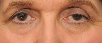
Puffiness under the eyes (photo 1)
Swelling can be eliminated using procedures that improve lymphatic drainage . These include vacuum diamond peeling or gas-liquid peeling jet peel. Treatment consists of a course of procedures, from 4 to 10 or more, depending on the manifestation and cause of the problem, if we are talking specifically about swelling caused by stagnation in the lower eyelid area.
Some formations that look like swelling are actually lower eyelid hernias . They are formed as a result of the redistribution and sliding of adipose tissue and a decrease in skin tone.
How to determine - edema or fatty hernia? If the size of the “bags” under the eyes varies: in the morning/evening, after the weekend/on weekdays, then most likely it is swelling.
Mesotherapy can also be used for swelling. Various meso-cocktails can strengthen the skin and remove dark circles, but a course of treatment is needed, since the effect of mesotherapy is cumulative. The course includes 3-5 procedures.
Hernia of the lower eyelid (photo 2,3)
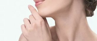
Small hernias, as in photo 2, can be corrected using contour plastic surgery, provided that the problem is not advanced. The area of the nasolacrimal groove is filled with hyaluronic acid filler. The bags are “leveled out”, unevenness and wrinkles are smoothed out almost immediately, and the effect lasts 10-12 months. There is no rehabilitation period!
Large hernias (photo 3) often cannot be repaired using non-surgical methods. If the formation under the eyes “hangs” strongly and is a complete semicircle, from the medial to the lateral edge, then only plastic surgery will help.
During surgery, fat packets are removed or redistributed to make them less noticeable. The operation is not a pleasant thing, but it gives 100% results for a long time.
After surgery, you can recover for weeks. Therefore, it is better to prevent such situations in time. To do this, we recommend undergoing preventive procedures to strengthen the skin of the eyelids after 30 years.
Nasolacrimal groove (photo 4.5)
The deep furrow under the eyes is smoothed out using a combination of contouring with fillers and mesotherapy.
If the problem is advanced, then it can only be dealt with through comprehensive correction. These are fillers + laser correction + mesotherapy. Additionally, IPL may be required - a technique of so-called photorejuvenation. You and I will have to “sweat”, but if we do not eliminate the problem at this stage, then it will only get worse.
Symptoms of eyelid lesions
Particularly common symptoms:
- Swelling and redness are characteristic of allergic reactions;
- Itching and inflammation of the skin - with infectious blepharitis;
- Swelling and/or pain along the edge of the eyelid, accompanied by redness - with barley (inflammation of the sebaceous gland), as well as inflammation with obstruction of the ducts of the meibomian glands, usually of the upper eyelid (chalazion).
When the position of the eyelids changes due to nervous disorders after cerebrovascular accidents, burns or facial injuries, the eyelids may not close. The eyelid, usually the lower one, can be in an inverted position, exposing the mucous membrane, as well as the eyeball. This condition is called ectropion. In this case, it is impossible to properly close the palpebral fissure, which leads to drying out of the conjunctiva of the eyeball, or even the cornea, and sometimes constant lacrimation is observed. Entropion of the eyelid is also characterized by the inability to completely close the eye slit. With it, the eyelashes are directed inward and, as a result, constantly irritate the conjunctiva, also causing lacrimation. Changes in the position or shape of the eyelids, their asymmetry, are often symptoms of underlying pathological processes in the eye orbit.
Symptoms of eyelid diseases
Symptoms of diseases of the eyelids are usually pronounced:
Swelling, redness and watery eyes most often indicate eye allergies.
Swelling of the edge of the eyelid, sometimes with local redness and pain - a sign of stye (inflammation of the sebaceous gland) or chalazion (inflammation with obstruction of the ducts of the meibomian glands)
Skin inflammation and itching are a symptom of blepharitis.
Among eyelid diseases, eyelid inversion is also distinguished. In this case, the eyelid is in a “screwed” state and exposes the mucous membrane of the eyeball. Due to improper closure of the palpebral fissure, the conjunctiva or even the cornea dries out, and the Patient usually experiences constant lacrimation.
Changes in the shape or incorrect position of the eyelids, as a rule, signal more hidden causes of pathological changes: cerebral circulatory disorders, nervous shock, and various injuries.
Treatment
Treatment of blepharitis with characteristic inflammation of the eyelids consists of prescribing antibacterial or antiallergic therapy.
A chalazion, especially one that tends to become frequently inflamed, should be excised surgically. However, it is worth considering that there are quite a lot of meibomian glands, and each of them, under predisposing conditions, can become inflamed.
Eversion and entropion of the eyelids are treated exclusively surgically, especially in cases of non-closure of the eye fissure, dystrophic changes in the conjunctiva or cornea, and incessant lacrimation.
Fomenko Natalia Ivanovna
Wrinkles and skin bags in the lower eyelid area
Laser resurfacing or laser blepharoplasty of the lower eyelids is a unique and very effective non-invasive procedure that helps with sagging skin of the lower eyelid (Fig. 6).
Non-surgical blepharoplasty (that is, eyelid lift) can only be performed with a laser. Other procedures (including those listed above) cannot be called blepharoplasty, since they do not in any way affect the area of eyelid skin .
Effect of laser resurfacing:
- will help eliminate skin ptosis;
- the eyelid will tighten, followed by the surrounding skin;
- wrinkles will smooth out or become less pronounced;
- the overall appearance of the skin will improve, dark circles will disappear;
- skin tone will increase.
In combination with injections, laser blepharoplasty works wonders. This can be seen in the photo: you can completely transform your face and maintain youth for a long time.
What definitely won't help?
Collagen injections, biorevitalization, various creams for bags under the eyes - all this cannot eliminate bags and even out the skin on the lower eyelid. The same applies to acid and chemical peels. They only help improve the condition of the skin, but they cannot tighten it or remove bags.
Trust the beauty of your eyes to our doctors and maintain your youth for the next 10 years!
Our trump cards : experience, education, licensed drugs and equipment and, of course, the results of our patients!
Carry out the procedure with a 15% discount!
We have many promotional offers, the main one: 15% discount on your first visit! We will select the best for you!
I want a discount!
Thank you!
Information has been sent to the clinic administrators!
Get a 15% discount on your first visit to our clinic! You can write to this number on WhatsApp!
The information in the article is of a general educational nature. It is important to understand that treatment cannot be prescribed over the Internet. It must be selected individually, based on a medical examination.
Therefore, sign up for a consultation with one of the doctors at our clinic, and then it will be clear in which direction you and I should move.
What to do if the upper or lower eyelid droops
There are non-surgical measures that will help with mild prolapse:
- Thoughtful eye makeup using predominantly light shades and giving the eyebrows the correct shape. These methods will not correct sagging eyelids, but will help make it less noticeable.
- Using cooling ice masks on the skin around the eyes. Such masks lead to some reduction in ptosis due to the narrowing of blood vessels and a reduction in tissue swelling.
- Healthy sleep , as well as sufficient hydration of the skin around the eyes.
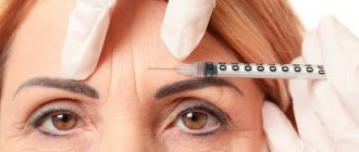
- Injection cosmetology with the introduction of botulinum toxins or fillers.


