There are literally only a few people who have not injured their nose at least once in their lives. This is due to the fact that the nose is the most prominent part of the face.
Author:
- Chuprikov Roman Sergeevich
ENT pathology expert
3.67 (Votes: 3)
There are literally only a few people who have not injured their nose at least once in their lives, since it is the most protruding part of the face.
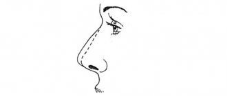
Features of surgery on the wings of the nose
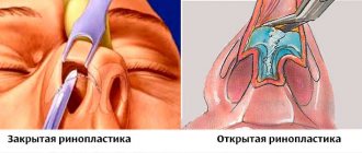
Correction of the wings of the nose can be performed as an independent operation or as part of a complex rhinoplasty.
If this is a complex operation, then the stage of nostril surgery is considered last, since some defects are eliminated in the previous stages. Depending on the scope of the intervention and the wishes of the patient, the method of anesthesia is selected. In the vast majority of cases, general anesthesia is used, but in certain situations local anesthesia may also be used. In any case, the patient will not feel pain, and our anesthesiologists will take care of his maximum safety and comfort. To reduce the size of the wings of the nose at its base, wedge-shaped incisions are made, excess tissue is excised, and the edges of the wound are sutured. If it is necessary to shorten the length of the wings of the nose, the incisions are made in a crescent or oval shape. Postoperative scars will be hidden in natural anatomical folds, which will make them invisible to others.
The narrowing of the wings of the nose is carried out by suturing the columella (skin septum located between the nostrils). Sutures are placed at its base, which, when tightened, narrow the width of the nostrils.
Recession of the wings of the nose is eliminated with the help of autografts - a piece of one's own cartilage tissue taken from another part of the patient's body, for example, from the ear.
Main function of the nose
The main function of this organ is respiratory, but aesthetic and communication functions will also be important. Scientific studies have been conducted that have revealed that people recognize each other by the shape and size of the nose, as well as by its proportions relative to other parts of the face. Thus, it is one of the most important signs of objective individuality.
Different races have their own characteristics of the structure of the external nose:
- Slavic - a relatively small bone part and a more developed cartilaginous section;
- Greek or Roman - distinguished by a long, narrow pyramid of the nose, often with a hump and a relatively small cartilaginous section;
- Caucasian - large bone and cartilaginous part, often with a hump;
- negroid - characterized by a small bony part and a large, usually flattened tip with wide wings;
- Asian - distinguished by proportionally small bone and cartilaginous parts, often with a flattened back and tip.
One of the features of our olfactory organ is that it grows throughout our lives, changing with age. This is especially true for the nasolabial angle. In children, the nasolabial angle is usually obtuse, about 105 degrees; in adults, it approaches a rectangular angle; in older people, the tip often drops and the angle becomes acute.
Preparation for the procedure
- The person being operated on must undergo a comprehensive medical examination and receive specialist advice.
- Before the operation, it is also necessary to visit a therapist, anesthesiologist and ENT specialist to select a set of medications and procedures.
- The surgeon performs a preoperative assessment of the appearance of the nose and takes photographs for further comparison.
- Modeling of the ideal size and shape of the nasal wings is carried out based on preoperative examinations.
- For several weeks before surgery, you should limit yourself to alcohol and smoking, not take medications that affect blood clotting, and not eat certain foods based on the advice of the specialist performing the operation.
Correction stages
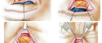
The first step is to make incisions in the side of the nose. The result of the entire operation depends on the depth and shape of the incision. Making incisions allows you to reduce the wings without affecting the internal cavity of the nose.
The second stage is removing excess tissue. The surgeon needs to be very careful here. If the doctor removes too much tissue, the aesthetic appearance may not be restored.
The third stage is connecting the skin using cosmetic sutures.
To reduce not only the wings, but also the nostrils, you need to remove a section in the inner cavity of the nose. The duration of plastic surgery ranges from 30-90 minutes.
Malakhov Alexander Andreevich plastic surgeon, specialist in rhinoplasty
How is the operation performed?
The content and method of carrying out the operation depend on the purpose for which it is carried out. Resection of the wings to narrow the base of the nose can be an independent operation, and in this case it is performed under local anesthesia. The surgeon makes an incision so that the postoperative scar is located in the natural groove between the wing of the nose and the cheek. After this, a strip of the wing of the nose adjacent to the cheek is removed (this part of the wing is devoid of cartilage, so only the skin and subcutaneous tissue are excised). The incision is carefully sutured, and a symmetrical operation is performed on the other side. Thinning of the nasal wing also involves tissue excision, but the skin remains in place, and only the subcutaneous connective tissue, which causes excess thickness, is removed.
As for the reconstruction and strengthening of the nasal wings, these procedures are usually performed as part of more extensive interventions that require anesthesia. A special case of such an operation is revision rhinoplasty. One type of iatrogenic (i.e. caused by a previous operation) deformity is retraction of the soft triangle. What is a soft triangle? This is a section of the wing adjacent to the columella and devoid of cartilaginous support (i.e., formed only by soft tissue). Due to its pliability, this area is easily deformed if the internal incision of the mucous membrane is too close to it, or if the suture stitch in this place is applied too wide. This deformity looks like a triangular retraction, causing an uneven contour of the nostril at the junction of the wing and columella and, unfortunately, is very persistent. Elimination of this defect requires local plastic surgery with the movement of mucosal flaps or the use of skin-cartilaginous grafts.
Finally, let's talk about surgical treatment of external new valve insufficiency. To strengthen the weak wing, thin long cartilage grafts from the own nasal septum, ear or rib cartilage are used. The added cartilage tissue increases the strength of the nasal wing and prevents the nostril from collapsing during breathing movements.
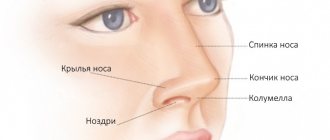
Anatomy of the external nose
The external nose has a complex anatomy, being an organ with a cartilaginous and bony skeleton, covered with thick skin. It consists of the following sections: the root (the area between the brow ridges), tapering downwards and turning into the pyramid - part of the bony skeleton of the nose, represented by the nasal bones and the frontal processes of the upper jaw.
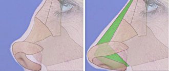
In the pyramid, there is a back - a face facing outwards, and slopes - the side surfaces of the pyramid. The tip of the nose is the most protruding part, represented by a cartilaginous skeleton. The tip consists of several cartilages: large and small alar cartilages, lateral cartilages (also called triangular) and quadrangular septal cartilage. All these cartilages are paired, with the exception of the quadrangular one. The cartilaginous section also includes: the wings of the nose, which form the respiratory openings, the nostrils and the collumella - the fold of skin between the nostrils.
The wings of the nose are covered from the inside with skin overgrown with dense bristly hairs; they form the vestibule of the nasal cavity, which passes into the cavity itself. The skin of the tip is very tightly fused with the underlying cartilage; in the bony part, the skin lies on loose subcutaneous tissue and is easily displaced. Between the skin and cartilage there are facial muscles, thanks to which we can wrinkle our nose, flare our wings, raise and lower the tip, which serves as a demonstration of emotions. The blood supply to the nose is very good. It comes from the system of external and internal carotid arteries. Venous outflow goes both to the internal jugular vein system and to the cranial cavity. Innervation is carried out by the branches of the trigeminal nerve.
Postoperative rehabilitation
Rhinoplasty in itself does not require special postoperative rehabilitation. Immediately after surgery, swelling develops in the intervention area caused by surgical trauma. The swelling subsides on its own within 10-14 days. Correction of the wings of the nose, as a rule, is not accompanied by the formation of bruises. External sutures, if any, are removed within 7-10 days. External scars are hidden in the natural furrows of the face, and are invisible in before and after photos a couple of months after the operation. In cases where alar plasty is part of a complex rhinoplasty, the content and timing of rehabilitation depend on the scope of the intervention performed.
Recovery after plastic surgery
Sutures placed during correction are usually removed on the sixth day after the intervention. The patient does not have to stay in the hospital all this time. You must remain under observation for the first few hours. A routine examination will take place just before the stitches are removed.
After the operation, tampons are placed in the patient's nasal passages, and a plaster bandage is placed on top to provide fixation. The scar will disappear only after 40-45 days. If corrected correctly, it should become completely invisible.
IMPORTANT! During the rehabilitation period, the patient should not visit baths, saunas and swimming pools, stay in the sun for a long time, put stress on the body and lift weights.
Sinusitis
Sinus inflammation usually occurs as a complication after viral and bacterial infections.
The main symptoms of sinusitis are headache and pain in the forehead and nose, prolonged discharge of yellowish or greenish mucus mixed with pus from the nose. Sinusitis requires mandatory diagnosis and treatment under the supervision of an otolaryngologist. Allow yourself to rest
Worried about a runny nose? The temperature has risen? Take a break from training. Infection carriers are not welcome in the gym.
Structure and function of the paranasal sinuses
The paranasal sinuses, connected to the nasal cavity, are peculiar air “caves” located in the adjacent bones of the skull. They, like the nasal cavity, are lined with mucous membrane. The main function of the paranasal sinuses is to lighten the weight of the skull bones, but they also serve as resonators for voice production. For a constant flow of air, all sinuses are connected to the nasal passages.
The largest of the sinuses, the maxillary sinus (also called the maxillary sinus), is located in the body of the upper jaw. Its bottom almost reaches the roots of the upper teeth, and the upper wall is also the lower wall of the orbit.
The frontal sinus is hidden in the thickness of the frontal bone (just above the bridge of the nose), its size can vary significantly. The thin posterior wall of this flat sinus separates it from the cranial cavity, directly behind it is the frontal lobe of the brain. Other paranasal sinuses (sphenoid and ethmoid cells) are also adjacent to the cranial cavity.
Diseases of the nasal mucosa
A runny nose is the most common disease in the world, which is why it is even included in the Guinness Book of Records. On average, an adult gets a runny nose up to ten times a year, and spends a total of up to three years with a stuffy nose throughout his life.
Nasal mucus, which has antiseptic properties, destroys a huge number of microbes trying to enter the body. If there are a lot of microbes, the volume of mucus also increases, which leads to a runny nose.
♦ Rhinitis. The mucous membrane of the nasal cavity performs numerous functions. However, its activity may be impaired. The most common cause of disruption of almost all functions of the mucous membrane is rhinitis (rhinos - nose, -itis - inflammation). Acute inflammation of the mucous membrane can develop as a result of hypothermia, exposure to a virus or mechanical irritant (dust, strong odor and even tobacco smoke). The mucous membrane swells, mucus secretion increases, breathing becomes difficult, and the sense of smell decreases. A person feels a tickling in the nose, complains of nasal congestion, headaches, thin transparent or thick greenish (if pyogenic bacteria are added) nasal discharge, sneezing.
Recovery will occur only after a few days, and if a person smokes or has a weakened immune system, then getting rid of the disease is much more difficult. There is a so-called vasomotor rhinitis, which develops as an allergic reaction to certain substances: dust, food, animal hair, etc. With this disease, nasal discharge is always liquid and transparent.
♦ Sinusitis. With colds, the infection can penetrate from the nasal cavity into the paranasal sinuses, causing inflammation. The cause of sinusitis (inflammation of the maxillary sinus) can also be dental pathology. When the sinuses are inflamed, they feel heaviness, severe nasal congestion, pain, and purulent nasal discharge is noted. Inflammation of the sinuses is dangerous due to its proximity to important organs such as the brain and eyeball. When the process is started, pus can destroy the bone walls of the sinuses, which can lead to catastrophic consequences.
Properties
Narrowing and expansion, the properties that wings possess, are achieved due to the presence of:
- dilator;
- transverse muscle;
- surface elevator;
- true dilator;
- septal depressor.
The area near the openings of the nasal openings is where the wings of the nose are located. Thanks to them, each person has an individual appearance. The formation of wings occurs in the womb, but after birth they are not expressed. The final form of this element is formed with maturation.
Reference! At about fifteen years of age, the wings are fully formed and no longer change.