Medical errors. Victoria Andryukhina (Andreeva)
Medical errors are possible in any field of medicine. In aesthetic surgery, if the doctor has performed unsatisfactory plastic surgery, as a rule, he performs free operations to correct the defect. But when a doctor does not want to admit his guilt, avoids responsibility, or, even worse, shifts it onto the patient, the only way out for the victim is to make a public statement.
Recently, popular blogger Lyubov Sidorkina (aka Lyubyatinka) on her YouTube channel shared her unsuccessful experience of contacting surgeon Victoria Valerievna Andryukhina.
Andryukhina Victoria Valerievna
- plastic surgeon, candidate of medical sciences. Victoria changed her last name, so on some sites she appears as Andreeva. For a long time she worked at the Vladimirsky MONIKI clinic. Today the place of work has changed.
Lyubov Sidorkina says:
“I asked about correction of two scars on my stomach. The first one formed in childhood after removal of appendicitis, the second - during emergency surgery in youth. As a result, a scar formed on the abdomen, which pulled the skin together to form a fold. The scar created an additional aesthetic defect: even with severe weight loss, the area of skin above the seam seemed to hang over and protrude, which created a bulge under tight-fitting clothing.”
According to the patient, she fell for the stories of the surgeon’s PR manager, who introduced Andryukhina as a famous and successful doctor. Lyubov came to Andryukhina with a scar problem, but to correct it, the doctor later chose the method of liposuction of the lower abdomen.
“Everything happened quickly. I was scheduled for surgery next week. After the operation, when I recovered from the severe pain, the first thing that caught my eye was the huge hematomas on my thighs in the groin area. Victoria warned that at first the stomach would look bruised. But he continued to look disgusting. I called the doctor and complained that my stomach looked strange. I would like to note that the initiative for interaction came only from me. The doctor reassured me and said that recovery could take up to a year. As a result, after six months it dawned on me that nothing would change and this was a medical error.”
Typically, in such cases, patients can sue the doctor. But Love did not do this, because she believes that it is useless. Before the operation, the patient signs a contract confirming that the doctor is not responsible for how the person’s body reacts to the operation.
Lyubov Sidorkina says that when she posted her post about Victoria Andryukhina on Instagram https://www.instagram.com/lubyatinka/ (post dated December 19, 2019), many people wrote to her in Direct and told her how this doctor ruined their appearance. One girl wrote that she filed a lawsuit, collected papers, and ordered an independent examination. The doctor did not appear at the first meeting, but only sent a representative. As a result, the patient lost the trial.
YouTube channel of Lyubov Sidorkina (aka Lyubyatinka) https://www.youtube.com/user/lyubaleyla/
MORE ABOUT LIPOSUCTION + TESTED CLINICS and SPECIALISTS performing liposuction!
Method of surgical treatment of rhinophyma
The invention relates to medicine, namely to maxillofacial surgery, plastic, reconstructive surgery, otorhinolaryngology. Selective destruction of water-saturated components of rhinophyma tumor tissue is carried out using a waveguide with an oscillation frequency of 25 kHz and an oscillation amplitude of 100 to 120 μm. Fibrous adhesions of rhinophyma are excised at an oscillation frequency of 35-55 kHz and an oscillation amplitude of 100 to 120 µm to healthy tissues layer by layer. Coagulation of the surface components of rhinophyma is carried out with an argon plasma coagulator at a flow rate of 1-3 l/min until a scab forms. The method allows to increase the effectiveness of treatment due to minimal thermal damage to tissues and reduced blood loss, which avoids severe cicatricial deformities of soft tissues and internal structures of the nose. 1 note
The invention relates to medicine, namely to maxillofacial surgery, plastic, reconstructive surgery, otorhinolaryngology, and can be used to treat patients with rhinophyma.
Rhinophyma (wine nose, pineal nose) is an inflammatory disease of the skin of the nose, characterized by the proliferation of connective tissue, blood vessels and sebaceous glands. When the disease occurs, the face becomes covered with ulcers, nodules and abscesses, and becomes earthy-gray. The most affected area is the nose, which over time begins to resemble a huge purple or dark red growth. The disease is often chronic, sluggish, so the nose with rhinophyma slowly becomes deformed over several years, then the course of the disease accelerates sharply and the nose becomes lumpy, blue-purple or dark red, and sometimes even purple. This deformation, due to the peculiarities of the anatomical structure of the nose, leads not only to cosmetic, but also to functional disorders. In this group of patients, there is damage to both the dermis and hypodermis of the nose; excess pathologically overgrown tissue can block the nasal passages, making nasal breathing difficult, therefore the complexity of the anatomical structure of the nose must be taken into account when restoring normal nasal structures.
There are different methods of treating rhinophyma.
After surgery, relapses of the disease often occur. There is a known method for removing rhinophyma, which consists in making shallow skin incisions, maximum atraumatic peeling of the skin from the tumor to the borders with healthy tissues, with wider preservation of the base of the skin flaps, peeling the tumor exposed from the skin from the underlying tissues, with maximum preservation of the integrity of the base of the nose, removal of the tumor either in parts, or as a single conglomerate, hemostasis of the wound surface, covering it with a preserved skin flap and joining with sutures (Shaturov A.G. et al. “Eponymous directory of otorhinological operations”, Irkutsk, 1984, p. 176).
This method is quite complex, traumatic and does not exclude the possibility of relapse of the disease.
There is a known method of surgical treatment of rhinophyma using an anterior vestibular incision of the skin of the nose. The skin of the nose is separated from the underlying tissues from the alar cartilages of the nose, cartilaginous and bone parts of the nose. Subcutaneous excision of the altered newly formed tissue is performed using high-intensity laser radiation. Afterwards, the wound surface is covered with a preserved skin flap and sutures are applied (RF Patent No. 2272589, IPC A61B 17/24, publ. 2005).
Despite the radical nature of the method, the disadvantage of this method is that the laser excises both changed areas and can also affect healthy fragments of cartilage and connective tissue, thereby causing inflammatory changes in the above tissues and subsequently leading to severe scarring. In addition, in this method, the skin of the nose is separated from the underlying tissues using a sharp method (scissors, scalpel), which leads to additional trauma and bleeding in the nasal area, as well as an increase in healing time and the patient’s hospital stay.
There is a known method of treating rhinophyma, which includes excision of altered newly formed tissue under local anesthesia, and the rhinophymatous nodes are first excised with an electrode in the form of a stretched string in the tip of the “Parus” electrodestruction apparatus “Fotek-80”, then hypertrophied areas of skin are cut layer by layer to healthy tissue, which evenly cover amorphous powder “Bismuth subgallate” to form a layer 1-3 cm thick on the surface. After the operation, Belanten ointment is applied and the drug Phlebodia is administered (RF Patent No. 2400164, IPC A61B 17/24, publ. 2010).
The disadvantage of this method is the external effect on tissues altered by rhinophyma; the internal rhinophymatous nodes are not affected, which can lead to the development of relapse. In addition, the method can lead to the development of severe scarring of the outer tissues of the nose.
The closest is the method of surgical treatment of rhinophyma, including excision of tissues of pathologically changed skin, coagulation of the surgical layer in the “spray” mode (RF Patent No. 2417776, IPC A61B 18/12, publ. 2011). This method uses argon-enhanced plasma coagulation of the surgical field using the Fotek EA140 device, first in the “fulguration” mode and then in the “spray” mode. In this case, tissue excision is carried out using a high-frequency loop electrode of the Fotek E80 device.
The disadvantage of this method is the superficial effect of the loop electrode on the tumor tissue, which does not allow radical removal of areas of rhinophyma located in the deep layers of the dermis on the internal structures of the nose, which will subsequently lead to the development of relapse due to the possibility of continued growth of rhinophymatous nodes under the skin of the nose. In addition, the proposed regime of argon-enhanced plasma coagulation can lead to the development of gross scar deformation that is undesirable in aesthetic terms.
The objective of the present invention is to eliminate these disadvantages, increase the effectiveness of treatment due to minimal thermal damage to tissue, which will avoid additional gross cicatricial deformities of the soft tissues of the nose and reduce the length of stay of patients in the hospital.
For this purpose, in the method of surgical treatment of rhinophyma, including excision of tissues of pathologically changed skin, coagulation of the surgical layer in the “spray” mode, it is proposed to excision of tissues of pathologically changed skin using the ultrasound of a Sonoca 400 generator. Moreover, first, selective destruction of the water-saturated components of tumor tissue of rhinophyma is carried out using a waveguide with an oscillation frequency 25 kHz and an oscillation amplitude from 100 to 120 μm, and then fibrous adhesions of rhinophyma are excised at an oscillation frequency of 35-55 kHz and an oscillation amplitude from 100 to 120 μm to healthy tissue layer by layer. Coagulation of the surface components of rhinophyma with argon plasma is carried out with a coagulator at a flow rate of 1-3 l/min until a scab forms.
The mechanism of argon plasma removal of rhinophyma tissue leads to the actual evaporation of pathological cells. Since argon plasma energy does not heat up, thermal damage to tissue is minimal. In addition, the method differs in that the generator allows you to create an ultrasonic signal that leaves intact healthy tissues that have an unchanged structure (vessels, nerves, cartilage tissue), which avoids additional blood loss, trauma and does not lead to gross scarring. Also, with the help of the APC 300 argon plasma coagulator, tissue charring does not occur, and the underlying tissues are not damaged and perforation of the skin flap does not occur. In addition, edema does not form in the postoperative period, due to which the healing process is short.
The method is carried out as follows.
With the patient under endotracheal anesthesia in the supine position, an anterior vestibular incision is made along the columella with transition to the septum and alar cartilages of the nose. The skin of the nose is separated with the underlying tissues from the alar cartilages of the nose, cartilaginous and bone parts of the nose using a Sonoca 400 ultrasonic generator with a waveguide installed on it with an oscillation frequency.
The generator allows you to create an ultrasonic signal that is converted (piezoconstrictor principle) in the instrument into mechanical vibrations, while the cavitation effect causes a selective effect on weak, moisture-saturated structures (destruction) and leaves untouched tissues that have an unchanged structure (healthy tissues), as well as vessels, nerves, cartilage tissue, which avoids additional blood loss and trauma.
An ultrasonic generator is used to continuously remove subcutaneous fibrous-changed newly formed tissue impregnated with large vessels. The tissue is fixed under slight pressure, then ultrasound is activated with an oscillation frequency of 25 kHz and an oscillation amplitude that increases or decreases depending on the structure of the tissues affected in the range from 100 to 120 μm, while the tissue is not subject to traction. Excision of the altered tissue is carried out to healthy tissues gradually, as if layer by layer, starting from the inner layers with the minimum power of the ultrasonic dissector.
Depending on the degree and nature of the pathologically altered tissues of rhinophyma, a mode of action on rhinophymatous adhesions is chosen. For small changes and thickness of rhinophymatous adhesions, the recommended exposure mode is usually from 35 to 40 kHz.
For large rhinophymatous conglomerates, it is recommended to use the exposure range from 35 to 55 kHz. In this case, pathologically overgrown subcutaneous fibrous adhesions of rhinophyma are excised using the lowest oscillation frequency of the dissector - 35 kHz at the periphery of the tumor, where these adhesions are less pronounced. Then the oscillation frequency is successively increased as the ultrasonic dissector moves from the periphery to the center of the tumor, up to 55 kHz depending on changes in the thickness of rhinophymatous adhesions, with radical removal of pathological tissue.
This treatment mode is as gentle as possible on the skin of the nose, which avoids the formation of through defects and deformations of the nose and does not lead to functional impairment and perforation of the skin flap. At the same time, the amplitude of vibrations was also changed from 100 to 120 μm, which allows destruction of blood vessels and sebaceous glands and leads to fibrosis of connective tissues.
This stage consists of a completely subcutaneous effect on the pathologically altered tissue.
Ultrasonic removal of fibrotic tissue depends on the volume of the lesion and is performed before healthy tissue. During surgery, tissue bleeding is minimal. The wound is washed with aseptic solutions. Next, pathologically altered excess skin in the area of the tip and wings of the nose is excised using scissors and sutures are placed with atraumatic monofilament on the skin and nasal mucosa.
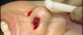
Then, using an argon plasma coagulator ERBE APC 300 at an argon flow rate of 1-3 l/min in the “spray” mode, the pathologically changed skin on the external nose is coagulated, a new shape of the nose is formed, and neighboring tissues are practically not affected, the vessels are instantly coagulated, which minimizes blood loss When using an argon plasma coagulator, tissue charring does not occur.
The proposed flow rate range provides the ability to select the rate of thermal tissue damage in the case of different stages of rhinophyma disease in the same patient in different areas of the nose (these patients have skin with ulcers, nodules and abscesses), which in turn leads to to a good cosmetic result in this group of patients. Those. we can vary this indicator depending on changes in the tissues in the nose area in order to avoid gross scarring in the area of the outer tissues of the nose.
Apply an aseptic bandage to the skin and secure with adhesive tape.
When using this method, blood loss is minimal, there is no swelling or various postoperative consequences, and the healing process is short. The sutures are removed 6-7 days after the operation. Rapid healing with this method leads to a good cosmetic effect and does not reduce the performance of patients.
Example.
Patient F., born in 1972, (case history No. 23329, 2011) was admitted to the maxillofacial surgery clinic with a diagnosis of Rhinophyma. She was clinically and laboratory examined. No contraindications to the operation were identified.
Under endotracheal anesthesia in the supine position, an anterior vestibular incision was made along the columella with transition to the septum and alar cartilages of the nose.
The skin of the nose was separated with the underlying tissues from the alar cartilages of the nose, cartilaginous and bone parts of the nose using a Sonoca 400 ultrasonic generator in continuous mode, i.e. We removed water-saturated components of rhinophyma tumor tissue to healthy tissue using a waveguide with an oscillation frequency of 25 kHz, while the amplitude of oscillations on the alar cartilages of the nose was 100 µm, and on the osteochondral part of the nose the amplitude was increased to 120 µm, i.e. the impact was carried out from the periphery to the center of the rhinophyma.
Using the method described above, pathologically overgrown subcutaneous fibrous adhesions of rhinophyma at the periphery of the tumor were excised with a dissector oscillation frequency of 35 kHz. Then the oscillation frequency was successively increased using 45, 55 kHz, while moving the ultrasonic dissector from the periphery to the center of the tumor, focusing on changes in the thickness of rhinophymatous adhesions, radically removing pathological tissue of the rhinophyma. In this case, the amplitude of oscillations was changed in the same way as when removing water-saturated components of the tumor.
After which the wound was washed with aseptic solutions.
Next, the pathologically altered excess skin in the area of the tip and wings of the nose was excised using scissors and sutures were placed with atraumatic monofilament on the skin and nasal mucosa. Then, using an argon plasma coagulator ERBE APC 300, argon-enhanced plasma coagulation was carried out at a flow rate of 1 l/min of pathologically altered skin on the external nose until a scab formed in the “spray” mode. An aseptic bandage was applied to the skin, which was fixed with an adhesive plaster.
Swelling and pain in the postoperative area were insignificant and resolved on their own on the 3rd day. The patient was discharged on the 4th day. At a follow-up examination after 10 months, there is no evidence of recurrence of the disease, aesthetically satisfactory scar tissue is determined, and a new shape of the nose has been formed, cosmetically satisfying the patient.
7 patients were treated using the proposed method. No complications were identified in any patient. Control examination of patients after 6-12 and 18 months did not reveal relapses of the disease.
The method, in comparison with known methods, allows to reduce treatment time, avoid additional gross scar deformations of both soft tissues and internal structures of the nose, which significantly improves the cosmetic result in this group of patients.
A method of surgical treatment of rhinophyma, including excision of tissues of pathologically altered skin, coagulation of the surgical layer in the “spray” mode, characterized in that the excision of tissues of pathologically altered skin is carried out using the ultrasound of a Sonoca 400 generator, and first selective destruction of the water-saturated components of the tumor tissue of rhinophyma is carried out with a waveguide with an oscillation frequency 25 kHz and an oscillation amplitude from 100 to 120 μm, and then fibrous adhesions of rhinophyma are excised at an oscillation frequency of 35-55 kHz and an oscillation amplitude from 100 to 120 μm to healthy tissue layer by layer, and coagulation is carried out on the surface components of rhinophyma with an argon plasma coagulator at a flow rate of 1- 3 l/min until scab formation.