There are 4 degrees of microtia:
Grade I:
A smaller version of the regular ear, with the same physical characteristics and the external auditory canal present.
Grade II:
Partially formed outer ear with a very small or narrow ear canal.
The ear canal may become closed (canal stenosis), resulting in hearing loss. III degree:
Absence of the outer ear of a typical shape (only a small remnant of cartilage and earlobe is present), absence of the external auditory canal and tympanic membrane (auricular atresia).
IV degree:
absence of ear (anotia).
Why does such a defect in appearance occur?
Doctors assign the main role in the appearance of anomalies in the anatomical structure of the ears to genetic causes. In addition, there are a number of provoking factors that lead to disruption of fetal development in the womb:
- infectious diseases of a pregnant woman (primarily of a viral nature, such as, for example, rubella);
- the expectant mother taking certain pharmacological drugs (for example, thalidomide);
- circulatory disorders of the uterus, placenta and fetus.
It must also be said that in more than half of the cases of microtia, the causes that caused it remain unclear.
Is microtia a genetic disease?
Yes and no.
No:
There are many families who have a child with microtia, but no one on either side of the family has ever had such a child. It appears to be a random event or anomaly occurring during early development. In the case of twins, one child may be born with microtia, but his twin brother will not.
Yes:
Microtia is observed in family generations. Sometimes, an aunt or uncle or cousin has microtia and a new family member is born with the same defect. Relatives may have the right ear affected, and then another family member may be born with a defect in the left ear. Or one family member may have bilateral microtia while the rest have unilateral microtia.
Laser otoplasty
In recent years, laser plastic surgery has become increasingly popular. It is usually performed to eliminate protruding ears. Instead of a scalpel, a laser is used, with the help of which all necessary manipulations are performed.
Advantages of this approach:
- The laser makes a thinner and smoother cut, which promotes rapid tissue regeneration
- There is no bleeding, since coagulation of blood vessels is carried out simultaneously
- Laser otoplasty is less likely to lead to infectious complications because the laser destroys bacteria in the wound
- Less risk of damage to blood vessels, nerves and other tissues
- Laser ear surgery takes just 30-60 minutes and usually does not require general anesthesia.
- Minimal risk of noticeable scarring
In view of these advantages, laser ear correction is gradually replacing traditional scalpel surgery. But still its capabilities are limited. In some clinical situations it cannot be used. The price of laser otoplasty is often higher than conventional surgery.
How will microtia affect my baby?
Microtia affects only external hearing.
Microtia may be accompanied by atresia (absence of the ear canal and often hearing loss), hemifacial microsomia, or other syndromes or genetic diseases such as Goldenhar syndrome or Treacher Collins syndrome. However, microtia does not affect a child's intelligence, motor skills, or success in life. Many parents of children with microtia are shocked at first, but soon realize how lucky they are because there are other children in our world who are born with life-threatening conditions. Microtia affects only the ear itself. Children can have different attitudes towards their ears, some don’t mind such ears, others worry. Try to encourage your child on those days when someone teases him about his ear, and the baby doubts his own confidence and feels upset. Help your child understand that he is no different from other children and can get whatever he wants from life.
Treatment
Treatment has two main goals:
- restoration of hearing, if necessary;
- elimination of a cosmetic defect.
Watch the facelift video - Non-surgical SMAS lifting using the Ulthera System device.
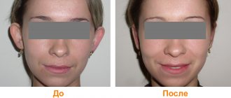
See photos before and after laser otoplasty in this article.
Surgical options for ear reconstruction for microtia
1. Using your own costal cartilage.
Reconstructive surgery using rib cartilage has been performed since the 1920s. It is a three to four step procedure that involves the use of costal cartilage (rib graft) and a flap of skin to cover the ear (skin graft). Surgery time for each stage can vary from one hour to five hours.
Because a rib ear graft is made from living body tissue, the ear will grow with the person. The reconstructed ear will be slightly stiffer than a normal ear because the costal cartilage tissue is denser, although to the human eye the difference is almost undetectable. The new ear will feel pain, it will bleed when damaged, and it will heal just like the real one.
2. Medpor surgical technique.
In this case, instead of costal cartilage, frames made of porous Medpor polyethylene are used to reconstruct the auricle. This material has been used for craniomaxillofacial operations for more than forty years. Medpor surgery can be performed in one stage, the surgery time varies from eight to twelve hours depending on the complexity of the operation. The Medpor ear is more rigid than the ear with a rib graft. Blood vessels are integrated into the porous openings of the Medpor frame, allowing the reconstructed ear to feel pain, bleed, and heal.
What else should you know?
The same factors that disrupt the anatomical structure of the auricle can negatively affect the formation of other organs and tissues. Therefore, if microtia or anotia is present, special attention should be paid to searching for other developmental defects in the child. It can be:
- changes in the structures of the middle and/or inner ear;
- anomalies of bone tissue of the facial part of the skull;
- absence or asymmetry of the lower jaw;
- cleft lip (“cleft lip”) and/or bones of the upper palate (“cleft palate”)
- disorders of the anatomical structure of the urinary tract and kidneys, etc.
To get more information about ear reconstruction in a medical clinic and to schedule a consultation with our specialists, call in your region or nationwide number 8 800 550 6891.
The material was prepared in agreement with the doctor, medical professor Khalil Ibrahim Janter.
Ear prosthetics
An ear prosthesis is an excellent option that does not require complex surgery. The prosthetic ears look amazing and very real! They can be attached using magnets or glue. You can swim and sleep with dentures. If you decide to reconstruct your ear using an auricle prosthesis made of silicone materials, contact Istok Audio Med LLC
. We cooperate with leading medical institutes and clinics throughout Russia.
119526, Moscow, Vernadsky Ave., 105, bldg. 2. Tkachenko Oksana ext. 525, mobile, Maria Klimacheva ext. 555, mobile,
For a more specific conversation about your case, please send:
- Photo
- Computed Tomography Data
- A copy of the IPR (if there is a disability)
How much does otoplasty cost?
Patients who decide to undergo otoplasty are always interested in how much ear correction costs. There is no single fixed price. Otoplasty is performed in different ways. Each case of ear deformation or protruding ears is individual. Therefore, the price of ear surgery varies significantly.
In addition, the cost of plastic surgery for the ears depends on:
- The authority of the clinic and plastic surgeon
- Operation techniques
- Countries, and even cities in which you are undergoing surgery
If reconstructive plastic surgery on the ears is necessary, the price is usually higher. Plus, sometimes you have to do it in several stages. That is, several operations are carried out alternately. Their number will determine how much ear surgery costs.
Send a request for treatment
What else can you do?
Nothing to do.
You always have the choice to do nothing and accept your microtia ear as it is, to love your ear. In fact, some people with microtia love their small ears and highlight them with piercings and short hair. Some even believe that their small ears are a distinctive feature, something that makes them stand out from the crowd. Visit foreign sites where you will find a lot of useful information about microtia:
In Russia, active communication on microtia issues is conducted on the VKontakte network, the microtia group https://vk.com/microtia
The article was written using materials from the sites:
How to get otoplasty in Germany?
A considerable number of patients do not travel to Germany simply because they do not know how to organize a trip. Indeed, for most people this task is quite difficult. You need to find a suitable clinic, contact and agree on everything with its management, draw up the necessary documents, translate medical documentation into German. Already in Germany you need to get to the right city, to the right clinic, book a hotel, find an interpreter to communicate with the medical staff.
BookingHealth can take care of all these concerns. Our advantages:
- Full organization of travel for treatment, starting with choosing a clinic and doctor, ending with transporting the patient from the airport to the clinic and back
- Accompaniment and support throughout the entire period of stay in Germany
- Saving on medical services. Thanks to BookingHealth, the cost of ear correction will be significantly lower. Plus you will save on services that German clinics offer at a significantly higher price
- Good and inexpensive insurance that will cover all costs in case of complications during treatment and for 4 years after its completion
- Any agreements with the clinic administration. We will agree on reducing the waiting time for an appointment, accommodation of an accompanying person, organizing individual meals and will resolve any other issues
To take advantage of the benefits of cooperation with BookingHealth, leave a request on our website. We will contact you as soon as possible.
Degree of development of pathology
Determining the age for surgical intervention, as well as all the nuances of the procedure, directly depend on the degree of development of this anomaly. Experts distinguish three degrees of development of protruding ears in children:
1. At the first stage of development of protruding ears, the angle of the ears does not exceed 45 degrees. Visually, such a defect is not immediately noticeable, and it can be disguised by choosing a suitable hairstyle - this is especially true for girls. To quickly correct protruding ears of the first degree, removal of excess cartilage tissue in the area of the recess of the auricle is most often used.
2. The second degree is characterized by a fairly significant protrusion of the ears, and the angle between the auricle and the head can range from 45 to 90 degrees. This pathology can be eliminated by thinning the cartilage tissue of the antihelix and subsequent surgical formation of the ear.
3. In the third degree of development, protruding ears are extremely pronounced, since the angle between the auricle and the head is more than 90 degrees. The correction technique involves removing excess cartilage tissue and artificially creating an antihelix.
Indications for surgical intervention at an early age are the second and third degrees of development of protruding ears, and in the first degree, correction of the auricles in children is carried out upon reaching adulthood.
Rehabilitation period
After a week, the doctor will remove the bandage, and the compression tape will only need to be worn at night. In about 2–3 weeks, the sutures heal, minor swelling disappears, and you can forget about correction once and for all.
The percentage of complications after ear correction surgery is quite low and occurs in only 5 people per 1000 operations performed. In order to eliminate the risk of developing any complications, it is important to follow the doctor’s recommendations during the rehabilitation period. These recommendations include:
1. Wearing an elastic bandage for at least one and a half weeks (the exact period is determined individually). You also need to use the bandage at night for another 5-6 months. This is necessary for the normal formation of ear cartilage.
2. Prohibition of physical activity (gym classes, etc.), as well as heavy physical labor for a period of 6 months.
3. Ban on visiting the sauna, bathhouse, solarium or beach for 3-4 months.
4. Mandatory attendance at prescribed physiotherapeutic procedures, which will speed up healing.
5. Mandatory dressings - usually on days 1-3-5 after surgery, day 12 - removal of sutures.
FAQ
— How can you get rid of protruding ears in an infant?
To correct the defect at the age of up to six months, special silicone molds are used that press the ears to the head. If they are not available, you can use tight-fitting hats. However, it is worth noting that the use of these methods does not guarantee 100% elimination of the defect.
— At what age should ear correction surgery be performed?
By the age of 6–8 years, a child’s ears are 90% formed—it is at this age that it is advisable to undergo surgery to correct protruding ears.
With the modern development of plastic surgery, correcting protruding ears is not a problem. However, it must be remembered that the operation will need to be performed on both ears, even if the defect is expressed in only one.
Correcting protruding ears at an early age can save the child from possible psychological discomfort and, as a result, the formation of complexes. Parents should not worry about scars, since they will not be noticeable behind the ears and the marks will disappear over time. The result obtained after the operation will remain for life.
The First Children's Medical Center sees pediatric surgeons of the highest category. Sign up for a consultation on surgical treatment from 8.00 to 20.00. Reception is by appointment only.
Children's health means parents' peace of mind!
Make an appointment with a surgeon
Choose a doctor
Our leading specialists
All specialists
Sheludchenko Tatyana Petrovna
- Chief otorhinolaryngologist of the network
- ENT
- Leading doctor
- Doctor of Medical Sciences
- Doctor of the highest category
Asmanov Alan Ismailovich
- ENT surgeon
- ENT
- Candidate of Medical Sciences
Davydova Marina Gennadievna
- Head of the Department of Otorhinolaryngology
- ENT
- Candidate of Medical Sciences
- Doctor of the highest category
Pugacheva Elena Nikolaevna
- Pediatric ENT surgeon
- ENT
Sedinkin Alexey Anatolievich
- ENT
- Candidate of Medical Sciences
- Doctor of the highest category
Churakov Pavel Valentinovich
- ENT
Progress of the operation
The operation to correct the auricles in children proceeds as follows: the surgeon makes a small incision at the back of the auricle (taking into account the hairline and the course of the natural fold of the skin). After the incision is made, the doctor forms a new form of cartilage (if necessary, excess cartilage tissue is removed). The surgeon then applies internal sutures. The final stage of the operation is cosmetic stitches, which over time turn into a tiny scar, which is also invisible behind the ear.
After the procedure, the doctor applies rollers aimed at maintaining the newly created position of the cartilage, and a securing bandage or bandage is put on top. The patient will have to wear them for 6 months.


