Breast examination after mammoplasty is carried out for two purposes. The first is an assessment of the breast tissue and lymphatic system. The second is to check the condition of the implants and the body’s reaction to them.
Breast examination begins with a medical examination and palpation. After examination, the surgeon prescribes an ultrasound examination of the mammary glands. Breast ultrasound after mammoplasty has advantages over magnetic resonance imaging (MRI) and mammography.
Ultrasound is a reliable, safe research method with no contraindications. The cost of ultrasound is 4-5 times lower than MRI. The duration of ultrasound diagnostics is 15-20 minutes.
Examination of breasts with implants.
Many girls wonder how breast examination will proceed after breast augmentation. Will all examined breast areas be visible on the machines?
Every woman, of course, takes care of her health. And everyone knows that after 35 years you need to undergo a mammogram once a year. And even more so after breast augmentation. Fluorography is also carried out once a year.
We all want to be healthy and therefore girls who care about their future always undergo examinations on time.
So what will this look like? Do implants interfere with breast exams?
How to do an ultrasound after mammoplasty? How is fluorography performed with breast implants? CT and MRI after mammoplasty? Ultrasound after breast augmentation? We will help you figure it out.
We would like to note that the presence of breast implants does not affect the examination in any way; you can always establish an accurate diagnosis using one of the methods.
But of course, with the availability of modern technology for examining the breast after mammoplasty.
Modern clinics, as a rule, are equipped with the latest models of technology. When making an appointment at a clinic for an examination, a girl should clarify what devices are available, whether it is possible to conduct an examination in this clinic if she has breast implants, and of course, consult with a specialist to select the exact research method in an individual case.
And we will dispel existing myths about the impossibility of conducting an examination.
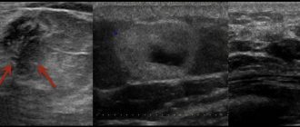
Ultrasound - ultrasound examination. After breast augmentation, it is performed annually. One of the most common examination methods in our time. It is also a mandatory examination before breast plastic surgery. Ultrasound of the mammary glands after breast augmentation allows you to identify pathologies of the mammary glands before surgery, assess the condition of the breast implants and the tissues themselves, and also exclude possible complications, such as inflammatory processes, tissue changes, and the formation of a capsule during the rehabilitation period.
Mammography after mammoplasty is the most thorough examination method. The mammography examination method after mammoplasty has minor difficulties. You need to know about this! The implant may block some areas of the mammary gland during the examination; to a greater extent and percentage, this applies to those cases when the implant is installed above the pectoral muscle. If the implant is installed under the muscle, the area of the breast covered is much smaller. Also, this research method is not informative in cases of ruptures or leakage of breast implants.
MRI after mammoplasty is a magnetic resonance imaging of the mammary glands.
A method for examining gland tissue using a powerful magnetic field. This method identifies tumor foci, metastases, and ruptures of breast implants.
CT or computed tomography after mammoplasty, this type is classified as X-ray methods for examining the breast. It is the most informative and accurate type of research for diagnosing cancer. CT scan is prescribed to clarify the diagnosis for a narrow circle of women.
FLG after mammoplasty or fluorography after breast augmentation.
Before undergoing this examination, the patient must notify the doctor about the presence of breast implants. Many people wonder whether implants are visible in the FLG image. We will also answer, yes, obviously.
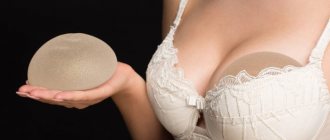
As you can see, the silicone implants that are used for mammoplasty are permeable to X-rays, their presence will not complicate the examination of the lungs during FLG.
Let's summarize.
To examine the breast after mammoplasty, examinations (ultrasound, CT, MRI) are carried out to obtain the most accurate data, and the presence of implants does not interfere with these examinations. And also examination of the lungs with FLH can also be carried out without fear.
Be healthy and beautiful.
46575 March 24, 2015 Author: My Plastic Surgeon Rate this publication: Rating: 3.7777777777778 based on 9 votes
Causes
Factors contributing to the development of contracture are not fully understood. However, it is known that they can be associated both with the characteristics of the operation performed and with various external reasons. The first group includes:
- Use of outdated models of endoprostheses (second or third generation).
- Mistakes when individually selecting an implant for a patient.
- Incorrect formation of the implantation pocket.
- Accumulation of blood in the implantation pocket (hematoma).
- Accumulation of serous fluid in the implantation pocket (seroma).
- Inflammatory process (suppuration).
- Failure to comply with the rules of asepsis and antiseptics.
- Violation of the integrity of the implant shell (rupture).
- Non-compliance with the regimen in the postoperative period.
- The body's predisposition to severe scarring (hypertrophic and keloid scars).
- Chronic diseases of the mammary glands.
Reasons not related to the operation include the individual characteristics of the patient’s body (for example, a tendency to form rough scars, even after minor injuries) and non-compliance with the doctor’s recommendations during the recovery period.
Absolute contraindications to CT
Contraindications to computed tomography are associated with radiation exposure to the body. They are usually divided into absolute and relative.
- The main and absolute contraindication to CT is pregnancy due to the negative effects of X-rays on the fetus.
- Children under 14 years of age are not recommended to have a CT scan without a referral from a treating specialist due to the risk of developing cancer from radiation.
Frequent X-ray examinations may be a limitation for computed tomography. The recommended radiation dose for medical purposes per year is 25 mSv. Exceeding this radiation exposure is dangerous for the patient's health.
MRI of the mammary glands
Breast MRI is the most accurate method for breast diagnosis. Unlike mammography, it does not have radiation exposure. However - just for prevention, for everyone - it is not suitable because:
- in St. Petersburg there are only a few specialists who can describe MRI of the mammary glands
- High-quality MRI of the mammary glands cannot be done on any MRI machine.
- The research is expensive, the state does not pay for it, and patients look for something cheaper.
Due to the fact that it is difficult to provide all these components, overdiagnosis during preventive MRI of the mammary glands is observed in all countries of the world.
NB! At the First Medical University named after. I.P. Pavlova at st. Lev Tolstoy 17 installed a mammograph with tomosynthesis function - CESM (3rd floor).
CESM is similar to MRI and is effective even in dense breasts.
Anyone can get tested on weekdays from 9:00 to 14:00 without an appointment. Contact the paid services department on the first floor. Cost from 2 (mammography without tomosynthesis) to 5 (CESM-tomosynthesis) tr.
If a tumor is detected, a consultation with an employee of the University Breast Center is free on the day of treatment.
What does a breast MRI show?
An experienced breast MRI specialist will always describe:
- tumor or tumors indicating its (their) size and location in the mammary gland: in which quadrant or on the border of which quadrants
- will indicate the distance from the tumor to the nipple in cm and/or mm, the distance from the tumor to the skin and muscle fascia - the doctor’s decision on the possibility of preserving the skin over the tumor depends on this
- will describe the condition of the skin above the tumor and in the area of the areola (whether there is swelling or not) - the doctor’s decision on the possibility of preserving the skin above the tumor or the need to remove the nipple and areola depends on this
- will describe the condition of the lymph nodes, but not only the axillary ones, but will also indicate the condition of the parasternal ones - which are hidden behind the cartilage of the ribs; will always compare them with the lymph nodes of the opposite side - as a sample of normal lymph nodes in a given patient - the doctor’s decision on whether you can perform a sentinel node biopsy or whether preliminary treatment is necessary depends on this
- will describe the condition of the bone tissue in the scanning area for metastases
- will provide information on the volume of breast tissue - for selecting an implant for one-stage reconstruction
- will provide information on the volume of the installed implant (if the passport for the implant is lost)
The accuracy of MRI is much higher if the images are evaluated “in dynamics” - new images are compared with previous ones. Therefore, when researching, always take the images on disk for your archive.
Hypervascularization
Malignant breast tumors on MRI specifically accumulate contrast due to their hypervascularization (increased vascular growth).
MRI diagnostics
Preventive MRI has shown better effectiveness only in combination with mammography in patients at risk for breast cancer caused by gene mutations (like Angelina Jolie). SEE ABOUT THIS .
MRI is also more justified in cases of dense breast tissue (as a rule, in the absence of childbirth and lactation), in case of small breasts without ptosis - when it is technically impossible to place them in a mammography machine.
The specialists of our Breast Center perform all types of plastic surgery on the mammary gland, including complications (ruptured implants, their displacement, etc.). SEE ABOUT THIS .
With which prostheses is it unacceptable to perform magnetic resonance diagnostics?
Although it is permissible to do an MRI with dental implants. During this examination, “internal” images of the body are obtained. A magnetic field is created in the tomograph, and the patient is placed in it.
The patient does not feel pain, and MRI does not damage tissues. The magnetic field created by the tomograph is 10 thousand times stronger than the Earth's magnetic field. It has the ability to attract objects that contain metal. If there are elements made of ferromagnetic materials in the magnetic resonance examination field, a potential threat arises. For this reason, people who have metal clamps on the aneurysm are not allowed to be diagnosed. The magnet can move the clamp - this will damage the vessel.
MRI diagnostics cannot be performed in the presence of the following types of implants:
- Cardio;
- Neuro;
- Ear;
- Containers that provide dosed dispensing of medications.
All electronic elements in the body, whose functioning can be disrupted under the influence of a magnetic field, the movement of which can create a health hazard - a limitation for MRI. But there are always exceptions, these include implants whose passport indicates their compatibility with the procedure.
How to behave after breast plastic surgery
When you wake up after the operation, put it on yourself (if you were not given compression garments in the operating room) or ask the staff to help you. Drains (tubes) are installed for several days, and even then - not after all plastic surgeries. They shouldn't scare you. They will be removed as soon as the amount of discharge decreases to 50 ml per day.
If you wake up and feel normal and feel the need to sit down, do not restrain yourself, sit down. If your head is not dizzy and you feel good, try to lower your legs down. If everything is fine, you can try to get back on your feet. If at one stage you feel dizzy, don’t force things - lie down and try again later.
You can drink and eat after you start sitting up on your own, provided that you really want to.
You can turn and lie on either side after surgery without restrictions. It is not advisable to actively move your arms. Also, do not force the time and do gymnastics for the first 2 days.
If any problems arise, call the nurse on duty.
We recommend that in the first days you do not refuse pain relief and do not endure pain (if any).
| For any problems, doubts or questions, do not hesitate to contact our staff. |
At home after the operation you will be somewhat limited, and at first you will probably need the help of your loved ones:
- Avoid heavy lifting and strenuous exercise for several weeks
- Do not put your hands behind your head (you will have difficulty washing your hair) after breast augmentation surgery with implants and mammary reduction
- You must stay active, do feasible work, and walk a lot
- Wear compression garments, no matter how uncomfortable they may be.
- Do not use (without consulting your doctor) lotions and creams in areas of post-operative wounds until they have healed.
- Do not wash in the bathtub, do not visit the pool or sauna until your doctor’s permission (at least 2 weeks)
- Check with your doctor for recommendations for scar correction (see HERE).
Treatment tactics
Capsular contracture is practically not amenable to conservative treatment. Only in grade 2 can botulinum toxin injections and local laser therapy be used. A positive effect is achieved in 30-45% of patients.
The third and fourth degrees of contracture require surgical correction in one of the following ways:
- Complete or partial excision of the thickened capsule.
- Re-endoprosthetics using textured implants.
- Formation of a new pocket for the endoprosthesis and reimplantation of already installed implants into this pocket.
The choice of surgical intervention tactics is determined individually. In some cases, simultaneous mastopexy (breast lift) is required to eliminate the deformity.
MRI for breast cancer
When describing, an experienced specialist will use the Bi-Rads system and avoid evasive terms and phrases with double interpretation. Also, he will always first describe the above points - the main thing, and then mention the less important - blood vessels and heart. Whereas a specialist “inexperienced in the mammary gland” will always first describe in detail what he knows better - the blood vessels and the heart. A conclusion will also be drawn up - an experienced one will first describe oncology, and then about blood vessels and so on.
An MRI of the mammary glands for cancer must be done before a biopsy. After it (due to injury), swelling, reaction of the lymph nodes, hematoma may appear: all this can falsely aggravate the signs of the disease.
THE algorithm for proper screening for breast cancer HERE .
Sometimes, when prescribing treatment for cancer patients before surgery (chemotherapy or hormone therapy, targeted treatment), an MRI of the mammary glands can also be performed to monitor its effectiveness.
The specialists of our Breast Center perform all types of operations on the mammary gland: for cancer and benign tumors, we can combine these operations with breast plastic surgery. SEE ABOUT THIS .