Defects and deformations of the auricle are very numerous and varied, both in shape and severity, and in the reasons for their occurrence. They can be either acquired as a result of mechanical, chemical or thermal trauma, tumor removal, or congenital - microtia and anotia.
…
Correction or complete restoration of the auricle after injury and, especially, as a result of abnormal development represent considerable difficulties and are one of the most difficult problems in plastic and maxillofacial surgery, despite modern advances in these areas.
Postoperative period
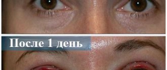
2-3 hours after otoplasty, the patient can go home with a pressure bandage. In the first days after surgery, there may be pain in the ear area. Therefore, immediately after otoplasty, the doctor gives detailed recommendations on taking painkillers and antibacterial drugs. The next day after the operation, doctors bandage and treat the wound. The bandage is replaced with a special pressure bandage (tennis tape), which the patient must wear constantly for 2 weeks.
Sutures are removed 8-10 days after surgery. The doctor conducts a follow-up examination after 1-2 months, by which time the swelling of the ears has completely subsided and the final result can be seen.
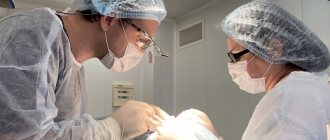
Medical otoplasty is performed by qualified doctors who have extensive experience in this field.
In addition, SM-Clinic has its own laboratory. This allows you to undergo preoperative examinations and pass the necessary tests in a short time.
Otoplasty, which is successfully performed by the plastic surgeons of our clinic, will allow you to get rid of a problem that has tormented you all your life in just one day.
| Author of the article: Makarov Andrey Vitalievich |
Otoplasty is a relatively simple operation that is performed under local anesthesia and under medicated sleep. It is performed on adults and children to eliminate a number of aesthetic and physiological disorders.
Complications after ear correction
- Subcutaneous hematoma is a fairly rare postoperative complication after ear correction. Its signs are quite characteristic: increasing pressing or throbbing pain, which is reported by the patient himself. Diagnosis is not difficult. Treatment consists of inspecting the wound, removing clots and finally stopping the bleeding.
- Epidermal blisters appear on the anterior surface of the auricle and along the edge of the helix. This type of complication occurs when there is excessive traumatic detachment of the skin from the cartilage, as well as the patient’s tendency to allergic reactions.
- Hypertrophic and keloid scars are more often observed after operations with excision of the skin of the postauricular fold and are rare in our practice, since we use gentle incisions with subsequent redistribution of excess skin.
- Ligature fistulas can develop with any type of intervention using internal permanent sutures. We use absorbable threads, which extremely rarely cause this type of complications.
- Recurrence of protrusion occurs when the degree of deformation and, consequently, methods for its correction are incorrectly assessed.
Anesthesia and duration of surgery
The operation is performed under local anesthesia or intravenous anesthesia. Its duration depends on the type and severity of the defect and takes from 40 minutes to 1.5 hours.
| In our clinic it is possible to correct several aesthetic imperfections at once in one operation, reducing recovery time. Simultaneous operations are the simultaneous performance of several surgical interventions on different parts of the body, for example, liposuction in parallel with correction of the shape of the ears. The surgeon will determine possible combinations of operations individually during the consultation. |
Correction of the ears for macrotia
Rice. 12
Abnormal enlargement of the auricle (macrotia) can be expressed either by an increase in the entire ear or an increase in its individual parts, and the increase is often due to the upper half of the auricle.
Rice.
13 Surgical correction of the auricles with macrotia consists of through excision of certain sections of it. In this case, the shape and location of the excised strips is directly dependent on the assigned correction tasks. So, for example, when the transverse size of the auricles decreases, crescent-shaped excisions along the helix are possible, and when the longitudinal size decreases, wedge-shaped excisions are possible.
However, in practice, the surgeon has to use combined incisions that reduce both sizes. This is due to the fact that the density of the shell tissue, caused primarily by cartilage, makes it difficult to bring the edges of the wound together during simple dissections without the formation of tissue bulges (“standing cones”).
Current trends in otoplasty
The prevalence of protruding pinnae is 5% in the Caucasian population, with the most common abnormality being flattened antihelix, followed by deep pinnae. Often these anomalies coexist, and both must be corrected.
More than 200 techniques for surgical correction of protruding ears have been described, suggesting that there is no single “best” method and that techniques will continue to evolve.
Otoplasty techniques can be divided into two broad categories:
- with cartilage removal
- with preservation of cartilage.
Cartilage cutting techniques aim to eliminate the inherent spring elasticity of cartilage and increase the stability (durability) of surgical results; These include cartilaginous incisions, wedge-shaped incisions, notching, and abrasion of the posterior or anterior surface of the auricle.
Cartilage preservation techniques have emerged in an attempt to prevent irregularities in the anterior surface of the auricle that can occur when cartilage cutting techniques are used.
In these cases, to create the desired contours of the auricle, sutures are used rather than cutting, which allows maintaining cartilage support and minimizing contour irregularities.
However, elements from both categories are often used together.
Historical methods, as well as more recent modifications, will be reviewed and discussed below.
Anatomy of the auricle
A thorough understanding of the anatomy of the normal and protruding ear is necessary for proper analysis before surgical correction.
The auricle consists of fibroelastic cartilage covered with thin skin. The skin of the anterior sections fits tightly to the perichondrium, but in the posterior section there is a layer of loose fatty tissue between the skin and cartilage.
The auricle consists of
- lobule,
- curl (helix),
- antihelix (antihelix),
- upper and lower legs of the antihelix (crus superior and inferior),
- triangular fossa,
- shell bowls (cavum conche),
- tragus, antitragus,
- intertragal notch (antitragus)
- The bowl of the sink is divided into two concavities. Several intrinsic and extrinsic muscles and ligaments stabilize the ear cartilage.
The blood supply to the auricle comes from branches of the external carotid artery and includes the superficial temporal, posterior auricular and occipital arteries. The posterior auricular, external jugular, superficial temporal and retromandibular veins provide venous drainage.
bones.
Sensory innervation occurs through the auriculotemporal nerve, the greater auricular nerve and the branches of cranial nerves VII, IX and X.
The growth of the auricle is 85% complete by the age of 3, and 90–95% of the adult ear size is achieved by the age of 5. The vertical length of the auricle is approximately 5–6 cm, and the width should be approximately 55% of the length. The long axis of the ear should be tilted back approximately 20°. The helix usually protrudes 1 to 2 cm from the mastoid skin, and the protrusion angle should be between 21 and 30°.
Timing of otoplasty
Protruding ears can be optimally corrected surgically at the age of 6-7 years, when they have almost completed their growth. Otoplasty at this age is performed to eliminate protruding ears before the child enters school in order to minimize ridicule from peers. The literature has documented significant psychosocial distress associated with the deformity, as well as improvements in many psychosocial parameters following otoplasty.
Performing otoplasty in children aged 6-7 years does not affect the further growth and development of the ear. An important advantage of performing otoplasty at a younger age is the increased compliance of the auricular cartilage, which allows for the effective use of cartilage preservation techniques and reduces the need for resection methods.
Purpose of otoplasty
The goal of otoplasty is an aesthetically acceptable restoration of the normal anatomical shape and anatomical position of the auricle.
The principles of ear surgery are as follows:
- The protrusion in the upper 1/3 of the ear should be eliminated.
- When viewed from the front, the helix of both ears should be visible behind the antihelix.
- The curl should have a smooth and regular line.
- The retroauricular groove should not be noticeably reduced or deformed.
- The ear should not be too close to the head - the posterior measurement from the outer edge of the helix to the mastoid process should be 10–12 mm at the top, 16–18 mm at the middle third and 20–22 mm at the bottom third
- The position of the two ears should match fairly accurately - within 3 mm at any point.
Treatment of protruding ears
Treatment for protruding ears includes non-surgical and surgical methods. Surgical methods can mainly be divided into two groups: cartilage cutting and preservation. These methods are often used in combination.
Non-surgical correction methods are described in detail at the link
Cartilage cutting methods
The first aesthetic otoplasties were examples of cartilage cutting techniques. Luckett noted the importance of reconstructing the underdeveloped antihelix fold in protruding ear surgery.
Cartilage dissection techniques were refined in the 1950s and 1960s by surgeons including Becker, Converse, Farrior, and Pitangi. These methods are most suitable for tough, thick cartilage that requires disruption of its inherent elasticity to maintain results.
The cartilage can be cut partially or full thickness from the front or back. Some of the cartilage may also be excised, scored, or rasped to reshape it.
When using any cartilage cutting technique, there is a risk of visible contour irregularities and sharp edges, which may lead to a failure to achieve the desired aesthetic result.
The technique described by Converse is an example of a classic but complex cartilage cutting technique. Parallel cartilage incisions are made through the full thickness of the cartilage along the edge of the concha and the desired antihelix fold to form an island of cartilage, which is then connected into a tube and secured with sutures to create a new antihelix fold.
The Becker technique also involves cutting through the entire cartilage.
Cartilage preservation methods
Cartilage preservation, or suturing, techniques have been developed to correct potential contour irregularities that may develop when cartilage cutting techniques are used.
Suture techniques are most suitable for thin or flexible cartilage. These methods do not create unevenness because the cartilage is not cut. However, in cases of stiffer cartilage, these methods may not provide adequate long-term correction because the repressive elasticity of the cartilage will remain intact.
Moustarger first described the use of multiple horizontal mattress sutures to reconstruct the antihelix fold in 1963.
In 1968, Furnas introduced the use of mattress sutures between the auricular cup and mastoid process to reduce protrusion of the deep concha cup.
After the patient is prepared and covered with sterile drapes, preoperative measurements are taken at three points: the upper pole, middle and lower third. The ruler is gently pressed against the scalp and measured from the outside of the helix at each of the above points.
These measurements are recorded by an assistant. The helix is then folded toward the mastoid process to visualize the line of the new antihelix fold, which is marked by a dotted line.
Retroauricular excision of the skin is carried out in the shape of an ellipse. And then Moustarget sutures are placed along the back surface at 3-4 points and Furnas.
Postoperative rehabilitation
Immediately after the operation, a special elastic bandage is applied for 5-7 days. Then it needs to be worn at night for 3-4 weeks.
Complications after otoplasty
Complications of otoplasty can be divided into early and late.
Early ones can occur within a few hours, these are hematoma, suppuration and cartilage necrosis.
Late complications occur over weeks to months and include suture extrusion, granuloma, recurrence or residual deformity, unacceptable scars (eg, keloid, hypertrophic)
Correction of earlobes
Rice. 14
Of the congenital anomalies of the earlobes, the most common are enlarged and split earlobes. Surgical reduction of the lobe can be performed together with reduction of the auricle, if there are indications for this, or in isolation. A simpler intervention is considered to involve excision of a wedge of tissue in the middle part of the lobe or near the cheek, resulting in a less noticeable postoperative scar.
The use of shaped incisions on the earlobes is due to the same reason as for corrections in other parts of the ear, i.e., the desire to prevent the formation of a “standing cone” of tissue at the ends of the incisions when stitching wounds.
Correction of the ears for penetrating defects
Among all the defects of the auricle, partial defects of the helix occupy a leading place. As a rule, such defects arise as a result of mechanical damage, or bites by a person or animal, less often due to thermal burns, electrical trauma, or removal of tumors.
Fig.15
To create a curl, flaps are used, cut on a wide base in the scalp against the curl defect. The flap is formed by two parallel incisions running from the scalp to the base of the auricle, where it is sutured to the posterior edge of the defect. This method eliminates the curl defect,