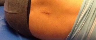
An umbilical hernia appears as a protrusion in the navel area that disappears or decreases in size when you lie down.
An umbilical hernia may be accompanied by discomfort, abdominal pain, and nausea.
The main cause of this disease is the weakening of the umbilical ring and the formation of a hole in the anterior abdominal wall, through which internal organs (intestines, greater omentum) emerge under the skin.
An umbilical hernia develops more often in women, which is associated with stretching of the umbilical ring during pregnancy.
Other causes of umbilical hernia include heavy lifting, chronic constipation, and obesity.
Why is it necessary to treat an umbilical hernia?
The very appearance of a hernia indicates that the organs have changed their location, and their normal mutual pressure on each other has been disrupted. This leads to disruption of the functions of all organs involved in the formation of a hernia.
An umbilical hernia must be treated. It cannot “drag out” on its own, and will only progress over time.
Without effective treatment, even a small hernia significantly impedes the normal functioning of organs
Lack of treatment will eventually lead to serious complications
- coprostasis - stagnation of feces in the large intestine,
- inflammation of the hernia - inflammation of the organs located in the hernial sac,
- strangulation of the umbilical hernia - sudden compression of the hernial contents in the hernial orifice, leading to disruption of the blood supply to the organs in the hernial sac, and as a result - necrosis (death) of tissue.
An umbilical hernia is often accompanied by chronic constipation. Constipation, in turn, leads to general intoxication of the body and can affect the condition of the entire gastrointestinal tract.
If a protrusion appears on the anterior abdominal wall, you should immediately consult a doctor, since it is much easier to treat a fresh hernia than one that has existed for a long time.
Postoperative hernias: general information
If simple hernias form primarily in physiologically weak places - along the white line, in the umbilical ring, inguinal and femoral canal, then postoperative ventral hernias - at the site of the scar, which can be under the right costal arch (after cholecystectomy) and under the left ( splenectomy), and in the iliac region on the right (appendectomy), above the pubis (after cesarean section), etc. Reasons for formation:
early:
- wound suppuration,
- peritonitis,
- failure of seams,
- increased intra-abdominal pressure,
- early activation of the patient;
late:
- lifting weights,
- flatulence,
- pregnancy,
- increased intra-abdominal pressure.
A hernia after abdominal surgery has a number of distinctive features:
- there is already a scar, that is, injury to the skin, muscles and aponeurosis (weak spot);
- possible adhesions inside the bag;
- the contents of the sac may become inflamed.
These points impose additional restrictions on the surgeon’s work and require the use of effective and modern treatment methods, especially at the stage of hernia repair.
Are there non-surgical methods for treating umbilical hernia?
The only way to remove an umbilical hernia is surgery. It is impossible to “set” a hernia once and for all without surgery.
The use of bandages in this situation also does not allow for a cure.
The use of new technologies, the use of modern plastic and suture materials, and many years of experience of our surgeons guarantee the highest quality of operations to remove an umbilical hernia, the absence of postoperative complications and relapse (recurrence of the disease).
Relapses
The probability of a reoccurrence of a hernia when using mesh endoprostheses is less than 10%.
Secondary hernia may occur due to:
- Incorrectly performed operation;
- The size of the mesh endoprosthesis is incorrectly selected;
- The mesh is incorrectly installed in the hernial orifice;
- The patient did not comply with all the requirements and instructions of the doctor regarding behavior and lifestyle in the postoperative period;
- Obesity;
- Prolonged cough;
- Constant constipation;
- Getting injured;
- Excessive physical activity.
Umbilical hernia surgery
Our clinic uses the most modern methods of treating hernias: obstructive hernioplasty using the latest 3D endoprostheses, open and endoscopic hernioplasty.
Hernia repair using endoprosthesis
Obstructive hernioplasty is the most effective surgical method for treating umbilical hernia. During the operation, the hole in the umbilical ring through which the hernia emerges is closed with a special multilayer mesh (endoprosthesis).
The foreign-made high-tech synthetic endoprostheses we use are very reliable, elastic, highly extensible and therefore do not limit the mobility of the abdominal wall.
The use of a mesh endoprosthesis protects the suture area from tension and thus provides three main advantages compared to the traditional surgical technique (tension plasty with local tissue):
- Extremely mild pain syndrome. Patients generally do not need to take pain medications after surgery.
- Short rehabilitation period. The patient goes home on his own the next day after the operation, and a month after the operation he can lift weights and play sports.
- Minimal risk of relapse. With proper placement of the endoprosthesis, recurrence of the hernia is practically impossible, whereas with the traditional technique it ranges from 6 to 14 percent.
The implant is not felt at all and does not cause any pain or discomfort.
Within a month after the operation, the mesh grows with connective tissue and over time, complete engraftment of the endoprosthesis occurs. The result is an anatomically unified complex that reliably closes the defect (weak spot) of the anterior abdominal wall and protects the tissue from repeated stretching.
We use two methods of installing an endoprosthesis: open and closed (endoscopic).
Open hernioplasty
With open hernioplasty, access to the contents of the hernia and the hernial orifice is through an external incision.
Next, hernia repair proceeds according to the following algorithm:
- isolation and opening of the sac with the contents of the hernia
- elimination of organ adhesions in the hernial sac, their reduction into the abdominal cavity
- removal of the hernial sac
- closing the hernial orifice using a special type of plastic surgery (hernioplasty)
- application and fastening of a special mesh implant of an individual shape
- formation of a cosmetic intradermal suture with a special suture material.
If the patient has diastasis (discrepancy) of the rectus abdominis muscles, which often accompanies an umbilical hernia, diastasis correction is always performed simultaneously with the operation to eliminate the hernia. This increases the effectiveness of the intervention and reduces the likelihood of disease relapse.
Our surgeons always perform the operation taking into account aesthetic requirements: the incisions made are minimal, the instruments used are atraumatic, and the sutures are applied using ultra-thin suture material.
Endoscopic hernioplasty
The most modern and low-traumatic method of removing a hernia is endoscopic, or closed hernioplasty.
This method is widely used in our clinic, since it has a number of undeniable advantages in the treatment of umbilical hernias:
- no visible scars,
- complete absence of pain syndrome,
- short recovery period (start of physical activity after a few days)
- the shortest rehabilitation period (100% return to active life in a maximum of two weeks)
- minimal number of relapses (less than 1%).
Unlike the classic open surgery technique, surgery is performed not through one large incision, but through three small punctures (0.5 - 0.6 cm).
Special endoscopic manipulators with a miniature video camera are inserted into them, sending an image to the monitor. With its help, the doctor monitors the progress of the operation.
The operation is carried out according to the same algorithm as with open access. But with endoscopic plastic surgery, a mesh implant is installed not through an external incision, but from inside the abdominal cavity at the site of the defect.
This method of treating hernias is carried out using special expensive multilayer meshes. One of the layers of such a mesh is made of a special chemical compound that prevents the formation of adhesions between the endoprosthesis and the abdominal organs.
Endoscopic hernioplasty gives better results because... the location of the mesh on the side of the abdominal cavity more reliably protects the defect of the abdominal wall with an increase in intra-abdominal pressure.
Endoscopic intervention eliminates the formation of postoperative hernias at puncture sites.
Abdominoplasty
If there are stretch marks, excess skin and subcutaneous fat on the abdomen, then it is recommended to combine the removal of an umbilical hernia with abdominoplasty.
This allows, simultaneously with hernia repair, to remove the skin-fat “apron”, eliminate sagging skin and stretch marks, and form a flat stomach and thin waist.
Surgery to eliminate an umbilical hernia can also be supplemented with liposuction of the abdomen or other parts.
Treatment
If after a medical examination a postoperative abdominal hernia is diagnosed, what should I do? The algorithm of actions depends on many factors - the size of the hernia, the presence of strangulation and the condition of nearby tissues, the general health of the patient.
The most radical and effective way to treat a postoperative hernia is to perform a repeat operation (hernioplasty) aimed at strengthening the abdominal wall in the area of the scar. This method will prevent relapses and eliminate possible complications in the future.
Types of hernioplasty surgery
Suturing the hernial opening
The simplest and fastest operation for eliminating a ventral hernia is suturing the defect by overlapping the muscles located along the edges of the hole. Before suturing, it is necessary to reduce the hernial contents into the abdominal cavity. The operation is performed only if the size of the hernial orifice does not exceed 5 cm, and there are no inflammatory or other pathological changes in the wound area. The method has a drawback - the possibility of a recurrence of a hernia in the same place (relapse).
Postoperative abdominal hernia, tension-free hernioplasty operation
The method is used in most cases of ventral hernias. The tissue in the area of the hernial opening is duplicated with a mesh implant. The material does not cause allergic reactions. An implant flap of the required size, like a patch, is sutured to the tissues around the hernia (muscles, aponeurosis). Due to its structure, the implant firmly fuses with the surrounding tissues and significantly increases the strength of the abdominal wall where there was previously a hernia. The use of a mesh allows you to avoid excessive tension of the abdominal wall in the area of the scar, which promotes better healing and avoids relapses in the future.
Laparoscopic hernioplasty
This is an operation in which surgeons bring a mesh implant to the hernia defect through 2-3 mini-incisions, just a couple of centimeters long.
This modification of hernioplasty has all the advantages of laparoscopy: rapid recovery of the patient after surgery, minimal scars after the intervention and low trauma. Qualified surgeons at our clinic will always correctly and adequately select the most optimal methods for treating a postoperative hernia or give detailed recommendations for treating a postoperative hernia without surgery. Such non-surgical methods are available to doctors and, in some cases, they can temporarily improve the patient’s condition and prevent complications and progression of the disease. But only hernioplasty surgery will help you cure a ventral hernia forever.
Treatment without surgery
If the doctor decides to postpone the hernioplasty operation for any reason, the following measures are recommended for patients to prevent the hernia from enlarging and complications:
- Wearing a bandage is a temporary measure. A bandage (a wide band of elastic fabric will relieve some of the load from the abdominal muscles) is applicable only in the initial stages of hernia development.
- Avoid physical activity, as it increases intra-abdominal pressure and increases the hernia defect and the size of the protrusion.
- Monitor bowel functions, avoid constipation and flatulence (bloating), as they also increase pressure inside the abdominal cavity.
Postoperative hernia: the diet provides for a limitation on the amount of food taken at the same time (you need to eat in small portions, but more often). You should not eat foods that cause increased gas formation - cabbage, legumes, fatty, spicy and spicy foods, carbonated and alcoholic drinks. You should also avoid foods that contribute to constipation - flour and bakery products made from premium flour, confectionery, saturated meat broths, rice, strong tea and coffee, astringent fruits and berries.
What is the most effective method for removing an umbilical hernia?
Our surgeons are fluent in endoscopic technology, but this technique is not always applicable for complex hernias. Often, much better results can be achieved by open hernioplasty.
Based on many years of experience in hernia repair, our surgeon will choose the optimal access method based on the characteristics of your particular disease.
The main factor in the successful outcome of hernia surgery is its impeccable technical execution. Poor surgical technique can ruin any method, even the best one. If all stages of the operation are performed correctly, then with any type of access the probability of hernia recurrence is minimal.
Before surgery
After consultation with one of our specialists regarding surgical treatment of a hernia and setting a preliminary date for hospitalization, the following tests must be taken:
- ECG (with description);
- Clinical blood test (with platelets, leukocyte formula, reticulocytes, ESR);
- blood test for group and Rh factor;
- blood test for sugar (glucose);
- prothrombin index + INR;
- APTT (kephalin-kaolin time), fibrinogen;
- Blood biochemistry (total bilirubin, direct bilirubin, AST, ALT, total protein, albumin, creatinine);
- Blood test for hepatitis B (Hbc antigen);
- Blood test for hepatitis C (anti-HCV antibodies total Ig M, Ig G);
- Syphilis blood test - antibodies to Treponema pallidum;
- Blood test for HIV - antibodies (Ig M, Ig G) to HIV types 1 and 2;
- General urine analysis.
Tests can be taken at the Phlebology Center at the address: st. 10th Anniversary of October, 9, st. Radio, no. 10, p. 9.
Make an appointment by phone
Before surgery, patients follow their usual daily routine and diet. The last meal before surgery is before 20-00. If you are worried, then at night you can take a light sleeping pill, calming herbal teas, and try to ensure 7-8 hours of sleep.
On the day of surgery, you should not wear makeup or metal jewelry. Do not eat or drink under any circumstances.
Hospitalization is carried out in the morning on the day of the operation (the time is set when registering for the operation). The clinic provides patients with: personal hygiene items, clothes and slippers for the ward, free 3 meals a day, a choice of dishes from the menu.
Umbilical hernia rehabilitation
Immediately after the operation, an elastic bandage is put on, which must be worn for a month.
At the BEAUTY DOCTOR clinic, patients are accommodated in single and double comfortable rooms.
The wards are equipped with continuous monitoring systems to monitor the patient's condition after surgery. Multifunctional beds create the opportunity to position and feed the patient after surgery in the most convenient position for him.
Each patient is provided with individual nursing care.
Since we use minimally invasive and maximally gentle techniques when repairing umbilical hernias, the postoperative period is easy and without any special complications.
You can get out of bed on the day of surgery. The next day after the operation, the patient goes home on his own, and after another 8-9 days comes for a follow-up examination and removal of sutures.
Two weeks after surgery, you are allowed to resume moderate physical activity (running, brisk walking). After endoscopic hernioplasty, such loads can be resumed within a few days.
A month after the operation, the patient can already lead a normal lifestyle and play sports.
How is the mesh used?
If a patient has a ventral hernia after abdominal surgery, implants can be used as follows:
- over the stitched aponeurosis without tension and opening the abdominal cavity for small defects;
- under the aponeurosis from the inside, between it and the peritoneum;
- between the leaves of the aponeurosis;
- combined hernioplasty using an implant and the patient’s own tissues.
The last two methods are widely used in our clinic, since in this case the implant takes on the main load and prevents recurrence of the hernia. In case of giant postoperative ventral hernias and tissue weakness, the mesh can be used for total prosthetics - modeling the anterior abdominal wall. In this case, the synthetic material actually replaces the muscles and aponeurosis.
Umbilical hernia surgery cost
| Umbilical hernia: hernioplasty | 32,000 rub. |
| Umbilical hernia: advanced hernioplasty | 37,000 rub. |
| Umbilical hernia: hernioplasty of recurrent hernia | 42,000 rub. |
| Umbilical hernia: endoscopic hernioplasty | 87,000 rub. |
| Umbilical hernia: hernioplasty with abdominoplasty | 260,000 190,000 rub. |
The cost of surgery to eliminate an umbilical hernia at the BEAUTY DOCTOR clinic includes all necessary examinations and dressings, as well as observation by a surgeon for six months after the operation.
Operations to remove an umbilical hernia are performed by highly qualified herniologist surgeons with extensive experience, trained in Russia and abroad:
| Gaboyan Aram Sergeevich, Doctor of Medical Sciences | Soboleva Polina Yurievna, Candidate of Medical Sciences | Malkarov Marat Azretovich, Candidate of Medical Sciences |
Surgery to remove a hernia using your own tissue
For small hernias (3 - 5 cm), the best treatment efficiency is shown by the method of suturing the hernial orifice using local tissue. During the operation, minimal tissue dissection is performed and the hernia opening is tightened with stitches. After this, the incision is closed and suture repair is performed, after which there are practically no visible traces of the operation. If there is a small layer of subcutaneous fat, surgery can be done using a surgical laser. The operation to remove a small hernia in adults is minimally invasive and does not require hospitalization; the complete recovery process does not exceed one month. Relapses after a well-performed operation are also excluded.
Do you want to get rid of a hernia quickly and permanently?
You're lucky to have found us. Contact us for advice.
- We will conduct an in-depth diagnosis of the condition of your abdominal cavity
- We will select for you the optimal treatment method from the entire range of modern high-tech surgical techniques
- Our highly qualified specialists - candidates and doctors of medical sciences - will perform the operation using the latest technologies, expensive specialized equipment and materials
- Your stomach will become healthy and beautiful, and traces of the intervention will be completely invisible to prying eyes
- We will carry out follow-up examinations and monitor your abdominal condition for six months to ensure there is no recurrence (free of charge)
How is implantation carried out?
There are 4 ways to install a mesh in the hernial orifice:
- Onlay . The mesh is sewn onto the aponeurosis of the rectus or external oblique abdominal muscles, directly contacting the subcutaneous fatty tissue.
- Inlay . The mesh is installed on the rectus or external oblique muscle, separated from the subcutaneous fat layer by the same aponeuroses.
- Underlay . The mesh is placed behind the muscle layer, usually separated from the abdominal cavity by the transversus abdominis muscle and transversalis fascia.
- Sublay . The mesh is located in the preperitoneal tissue, separated by the peritoneum from the abdominal organs.
The surgeon individually selects the appropriate implantation method for each patient.
In this case, it is necessary to follow the rules for installing mesh endoprostheses:
- the hernia mesh is installed only within the aponeurotic and muscle tissues;
- multifilament threads cannot be used to secure the endoprosthesis;
- For mixed truncation, a non-absorbable type of mesh endoprosthesis cannot be used.
Methods for diagnosing hernia
Diagnosis of a hernia is carried out during examination of the patient. In this case, the doctor uses the methods of palpation (palpation), percussion (tapping) and auscultation (listening to the natural sounds of the body).
To obtain a more complete picture, instrumental studies are performed:
Radiography
Radiography of a hernia allows one to obtain additional information about the presence of adhesions, parietal strangulation of the hernia and partial intestinal obstruction.
More information about the diagnostic method
Ultrasound examinations
Ultrasound makes it possible to clarify the location of the hernia, the shape and size of the hernial orifice, assess the condition of the surrounding tissues (this allows you to choose the most effective technique for reducing the hernia), and determine the contents of the cavity of the hernial sac.
More information about the diagnostic method
Computed tomography (CT)
Computed tomography for hernia is used if ultrasound data is insufficient.
More information about the diagnostic method
Sign up for diagnostics To accurately diagnose the disease, make an appointment with specialists from the Family Doctor network.