The laser device leaves behind small wounds on the skin, which crust over over the next few hours. Noticeable swelling and redness may also occur, which is a completely normal reaction to laser photocoagulation. Typically, minor damage resulting from laser mole removal goes away within 2-4 weeks. How long the wound will take to heal is influenced by the size of the removed lesion, the technical features of the procedure, and the patient’s compliance with the prescribed standards.
Removing a mole takes a couple of minutes, but complete restoration of the skin will take at least a month.
What are moles? What are they?
To answer this question, you must first clarify what we mean by the word “mole.” An ordinary person can easily find several types of skin formations. Brown, red, flesh-colored - all these can be called moles. In fact, the World Health Organization (WHO) classification of benign skin lesions includes about 150 names. And all this diversity can also be called moles. Read a little more about the most common types of moles here.
As we said above, there are a huge number of types of moles. Today we will talk about those that are most common and, theoretically, can pose the greatest danger - brown and flesh-colored moles.
How does mole burning work?
The procedure for removing a nevus with an electric knife includes the following steps:
- Visual inspection of the birthmark.
- Treating the surgical field with a skin antiseptic to prevent infection from entering the wound.
- Local application of a local anesthetic.
- The procedure for removing a nevus using an electric knife.
- Treatment of postoperative wound.
Burning out a nevus with an electric knife takes place in one movement, so you will not feel pain or burning. Some patients experience a slight tingling sensation where the device comes into contact with the skin.
Important! Self-removal of skin tumors is strictly prohibited. This can lead to the development of undesirable consequences such as inflammation, sepsis (blood poisoning), skin cancer. If necessary, you should contact a specialist at a medical clinic.
Brown moles
Such moles are a cluster of nevus cells. These cells are very similar to melanocytes and, like them, produce the pigment melanin. This pigment is brown in color, which makes our skin darker if there is a lot of it and lighter if there is not enough of it. Its production also increases after prolonged exposure to the sun.
Melanocytes and nevus cells
Melanocytes and nevus cells have certain differences, which we will not delve into. Let’s just note that brown moles most often consist of nevus cells. It is nevus cells that can turn into melanoma cells - one of the most malignant human tumors (more about it here)
Stages of healing
The first stage of healing lasts the first seven days, during which the crust gradually darkens and becomes more rigid. You should never touch it, as it protects the wound from infection and promotes the appearance of new healthy tissue. In the first week after the procedure, the operated area should be protected from any injuries and damage; it should not be covered with clothing too often, wetted with water or rubbed with a washcloth, scratched or applied with any cosmetics.
The use of disinfectants is allowed if they have been prescribed by a doctor. You can also treat the crust with a mild solution of potassium permanganate.
The next stage of healing includes the second week after surgery. During this time, the dark crust at the site of the former mole disappears on its own, and light pink skin remains instead. It is still recommended to protect this area from any excessive exposure as well as ultraviolet rays. To this end, it is worth reducing the time spent in the open sun, as well as treating the skin with sunscreen with a maximum protective factor (about SPF 50). Young skin can become significantly pigmented when exposed to the sun, especially on the face.
The final stage of recovery lasts 14 to 20 days after surgery or more. By this time, completely healthy skin has formed at the site of the removed mole, which has protective factors from ultraviolet radiation and other external influences, including mechanical ones. A slight itching may persist, which can be relieved with soothing ointments. Otherwise, skin surface care is no longer required.
If a hole remains at the site of the removed lesion, do not worry: usually the skin smoothes out on its own over the next 1-2 months.
Why can mole removal be dangerous?
If you have already read about moles on the Internet, then you have probably already found information that “after removing moles, you can die from cancer.” I discuss this topic in detail in this article. Now I will say that such cases really do happen. This happens when a doctor removes a malignant tumor, which he considered an ordinary mole, and does not send it for histological examination. After this, indeed, melanoma can very quickly develop at the site of removal and this can cause death.
“Will you take a test from the mole the day before?”
The only analysis that can reliably determine the nature of the formation is histological examination. The formation is removed entirely and sent for pathomorphological examination. No “plucking” is performed in advance before removing moles.
It is possible to perform a cytological examination in advance if there is discharge, ulceration or trauma to the formation. This study allows you to establish a preliminary diagnosis and is performed when it is difficult to make a preliminary diagnosis. Often, an external examination or dermatoscopy is sufficient to make a diagnosis. For example, in relation to fibroepithelial polyps (papillomas), keratomas, fibromas, viral warts, a fairly large group of nevi (intradermal, warty, non-pigmented, etc.).
Reasons for removing moles
In my opinion, there are 4 reasons to remove moles:
- The mole raises doubts about its benign quality among the oncologist. In this situation, we will not be talking about removing a mole, but about a biopsy of a possible malignant tumor. Subsequent histological examination will show whether a larger operation is needed or not.
- The mole is subject to regular trauma. This can happen when playing sports, shaving, during work, when a mole is located on the belt or bra line, as well as in a hairdresser. Regular traumatization of a mole, especially to the point of bleeding, may be one of the risk factors for the development of melanoma.
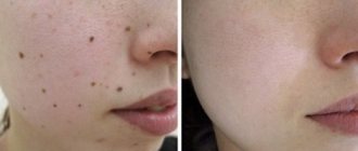
- A mole causes a cosmetic defect. No explanation needed here. The issue of beauty is very individual for each person and there are no clear rules here.
- A mole causes obsessive thoughts about its transformation into cancer or melanoma (oncophobia). As a rule, this happens after a long time of independently studying the Internet on the topic of melanoma and skin cancer. Sleep and appetite are disturbed, neurosis sets in, because if you want, you can find confirmation of anything on the Internet. Not only that “this mole I have is definitely melanoma,” but also that “Lenin is a mushroom.” In this situation, removal of the mole is necessary only to restore lost psychological comfort.
In the latter case, it is important to combine deletion with stopping independent study of the World Wide Web on the topic of skin cancer and melanoma.
Possibility of unforeseen consequences
Laser correction of moles is one of the safest ways to get rid of nevus, avoiding complications and unforeseen consequences. A beam of directed light radiation literally evaporates the pigmented cells of moles, which is visually manifested by the complete removal of the epidermal formation in just a few minutes. Minimization of negative consequences occurs due to the following objective factors.
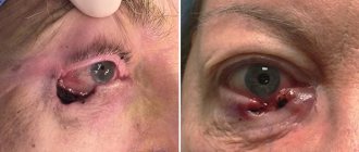
- The laser beam acts in a strictly targeted and selective manner, without injuring healthy skin structures, which allows the technique to be used even to remove the smallest nevi in the eyelid area and mucous membranes;
- There is virtually no risk of infection, since the laser exhibits a powerful antibacterial and antimicrobial effect, protecting the wound surface from infection;
- Laser destruction is not accompanied by bleeding or hemorrhage, since during the manipulation process all blood capillaries supplying the body of the mole undergo thermal coagulation;
- The laser activates regenerative processes in the skin, which significantly accelerates wound healing;
- The technique is low-traumatic, which avoids the formation of connective tissue and all the negative consequences associated with this factor.
Despite the safety of the method, it should be borne in mind that before the procedure it is necessary to carefully study the list of contraindications, since the consequences and complications of laser correction most often result from neglect of this list, which includes the following conditions:
- Pregnancy;
- Acute infectious process;
- Oncology of any localization;
- Inflammation in the intended area of laser exposure;
- Period of menstruation in women;
- Immunodeficiency;
- Exacerbation of chronic pathologies;
- Hyperthermia.
Contraindications to burning moles
New growths on the skin can be benign or malignant. Removing moles with a coagulator is considered safe provided that the nevus does not show signs of developing a cancerous tumor. The doctor determines the primary signs during a visual examination. You cannot remove a nevus using an electric knife if you suspect the formation of a malignant process. Also contraindications include:
- bleeding disorders
- intolerance to electrical procedures
- allergic reactions to local anesthetics
- herpetic infection in the acute phase
- an inflammatory skin disease at the site where a mole was burned out with an electric knife
Any such case must be eliminated before electrocoagulation is performed.
I monitor one mole, remove another: how not to miss dangerous neoplasms on your skin
Where do they come from
On the Internet you can find a version that our moles initially grow... due to tanning. They say that a person is born with clear skin, and over time it is because of the sun that moles appear on it.
“In fact, the formation of moles and their number is determined primarily by heredity,” says dermato-oncologist at the Clinic of Skin Diseases, assistant at the Department of Skin Diseases at Sechenov University, candidate of medical sciences Ekaterina Vertieva.
- That is, those who have a lot of new growths on the skin in their family - mom, dad, grandmother or grandfather - will have more moles.
The second factor is the sun. Under the influence of its rays, the number of moles increases.
At the same time, there are several peaks in a person’s life when most moles appear. Such surges are associated with hormonal changes: at a younger age (3 - 6 years), adolescence and from 25 to 35 years. Women may experience additional peaks during pregnancy.
— Is it true that there is a “safe limit” of moles, and if there are more than normal, then a person has an increased risk of skin cancer?
— If there are more than a hundred moles on the skin, then the risk of malignant neoplasms is really higher. But not so much that those with a rich scattering of moles should immediately sound the alarm. The logic is simple: the more tumors there are, in principle, the higher the likelihood that among them there will be nevi with malignant potential (how to protect yourself from danger - see below).
STAY IN TOUCH
What is what
Among the names of growths on the skin you can find the classic, familiar word “mole,” as well as nevus and keratoma. What is the difference?
“ Nevus is the medical name for the same mole,” explains Ekaterina Vertieva. - Essentially, this is a benign skin tumor. A mole consists of pigment cells of melanocytes, which produce the coloring pigment melanin (because of it, our skin darkens when tanning. - Ed.) A mole is a cluster of such melanocyte cells. That is why the appearance of nevi is associated, among other things, with exposure to the sun.
— Keratoma is a benign superficial formation on the skin that can never degenerate into something malignant. Keratomas occur more often in old age. They consist of keratinocyte cells (these are the surface cells of the upper layer of the skin of the epidermis). Externally, a keratome usually looks like a brown formation rising above the surface of the skin. Keratomas often become crusty and flaky. Pieces can fall off, which scares people. In fact, this is a natural desquamation process. There is no need to be scared, the doctor reassures.
On a stem and with hairs - what can you expect from them?
- There are moles that seem to move away from the skin and rise above it - is this dangerous?
- Look: we have superficial layers of skin, the epidermis, and deep layers, the dermis. Common, classic dark moles are superficial, so-called border nevi. They are found in the epidermis. Over time, some superficial nevi absolutely physiologically, that is, safely, naturally grow into the deep layers of the skin, the dermis. And then they begin to rise above the surface of the skin. Nothing wrong with that.
Moreover, over time, by about 50-60 years, dermal nevi completely lose their pigment. They cease to be brown and acquire the color of healthy skin. This is the so-called mole involution. All this is absolutely safe.
- When do hairs grow from a mole? They say this is a good sign, such nevi are definitely safe and will not degenerate into melanoma.
- This is a misconception. Melanoma can easily grow hair. Therefore, if a nevus has suspicious signs (see below), then the hairs are not a reason to calm down and not check the mole.
By the way, doctors categorically do not advise pulling hair out of a nevus; this is unnecessary trauma. You can only cut it carefully.
Meyerson phenomenon
This is the name of the condition when redness appears around the mole, like an inflamed pink corolla, our expert explains. And it reassures: as a rule, it is not dangerous. Most often, the Meyerson phenomenon occurs in patients with a tendency to skin diseases: eczema, atopic dermatitis. If you experience any discomfort or itching, you should consult a doctor. Usually, dermatologists prescribe atopic corticosteroids to relieve such symptoms; with their help, the symptoms are easily removed.
— If you damage a mole and it bleeds, do you need to see a doctor urgently?
— It is necessary, but not necessarily right on the same day or the next day, up to 7 days is quite normal. There is no need to be afraid that after injury, even with bleeding, your mole will definitely degenerate into something bad. Do not believe the stories about a grandmother who picked off a mole, it immediately metastasized into the blood, and death quickly occurred. It is a myth. If a person injures a benign mole, then nothing bad will happen to it. Adverse consequences occur if it was initially melanoma.
— What to do immediately after a mole is damaged, before going to the doctor?
- Treat with a clear antiseptic. Hydrogen peroxide, for example. Just don’t smear it with fucorcin, iodine or brilliant green. Because it will then be difficult for the doctor to examine and assess the condition of your mole due to staining with these substances.
How often to check with a doctor
There are tips on the Internet to show your moles to a doctor every 6 to 12 months.
“If a person has 100 or more nevi and is advised to visit a doctor every six months or a year, then this is still too much,” says Ekaterina Vertieva. - In most cases, moles will not turn into anything bad. But, to be sure of this, it is important to undergo a full examination once by a dermatologist, or even better, an oncodermatologist. Have a specialist carefully check all moles and confirm that none of them are cause for concern. Or he indicated those that could potentially be dangerous, and then told how often they need to be monitored.
If the doctor, after examination, says that all moles are calm, then, as a rule, it is recommended to see the next time in 3 - 4 years. At the same time, no one canceled the self-examination.
THIS WILL BE USEFUL
The golden rule for self-examination
This is the international standard for assessing moles. It can be used as a test at home. The mole is tested according to the ABCDE criteria.
A (asymmetry, asymmetry) - has the mole become asymmetrical?
B (border) - have the borders of the mole become uneven?
C (colour) - has the color of the mole changed: several shades of brown, black, blue, pink have appeared?
D (diameter, diameter) - has the mole grown more than 6 mm in diameter?
E (evolution, evolution) - does the mole change intensively over 4 - 6 months (in shape, color, size)?
Important : if the answers to all questions are “yes,” this is reason to suspect the mole is malignant and immediately consult a doctor. If you have several of the symptoms, then the risk is lower, but you also need to see a doctor.
Delete cannot be saved
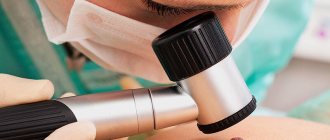
“There are three reasons for removing moles,” says Dr. Vertieva. — Cosmetic removal — if a person simply doesn’t like the way a mole looks. We also recommend removing benign moles if they are located in places where they are often injured. For example, for men in the beard area, for women in the bra clasp area. The third option is if we see suspicious symptoms. In these cases, the doctor conducts an examination using a dermatoscope (a device similar to a microscope. - Ed.). If a mole is causing concern, removal is done for medical reasons.
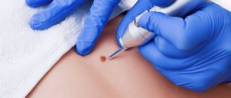
— What are the current methods for removing moles?
- Laser, radio wave method, electrocoagulation (cauterization of the nevus with high-frequency current) are used - these methods destroy the mole. As well as surgical excision.
If the patient has a suspicious nevus, then surgical removal is necessary. Only in this case, the tumor tissue is fully preserved, and histologists will be able to give a reliable conclusion as to whether there are malignant cells in the mole.
In other cases, when the mole is definitely calm, the least traumatic method is the radio wave method or laser (coagulation is a rougher method). After removing the nevus, a neat wound remains; it must be treated with a solution of potassium permanganate or fucorcin. A crust forms, which falls off after an average of two weeks. You can wet the wound site with water from 3 to 4 days.
I strongly do not recommend self-medicating at home and using celandine to remove tumors. At best, this technique leads to a chemical burn, and sometimes to more serious consequences in the case of malignant potential of the formation.
QUESTION ON THE TOPIC
Is it possible to sunbathe if you have a lot of moles?
“Yes, this is not a contraindication for tanning,” our expert rejoices. — The main thing is to follow safety rules: stay in the sun before 11 a.m. and after 4 p.m. And use sunscreen.
BREAKTHROUGH AND PROSPECTS
“Now there is a real revolution in the treatment of melanoma, the most aggressive skin cancer,” says Ekaterina Vertieva. — Previously, chemotherapy was used, which was very toxic and at the same time extended the life of patients by only a couple of months. Since the beginning of the 2010s, targeted drugs have been used. They do not hit all cells of the body, like chemotherapy, but selectively hit specific targets. These drugs are available in Russia under compulsory medical insurance and prolong the life of our patients very significantly. The main trend now is the study of new targets for the fight against melanoma. And also immunotherapy (the method for which the Nobel Prize in Medicine was awarded this year. - Ed.).
CONGRATULATIONS!
The oldest department of Sechenov University is 150 years old
On May 27, the Department of Skin and Venereal Diseases named after. V.A. Rakhmanov Sechenov University - 150 years. Already in the first years of its work, the department was recognized as the best in Europe. Over a century and a half history, vast experience in teaching the science of dermatovenereology has been accumulated here. Today, about 2,500 skin and venereal diseases are known, say department employees. The most common are psoriasis, atopic dermatitis, eczema and fungal infections; In recent decades, there has been a steady increase in the incidence of allergic dermatoses.
The head of the department, Professor Olga Olisova, lists modern promising scientific developments of the department's employees: improving the early diagnosis of lymphoproliferative skin diseases (T-cell lymphomas) using genetic markers; development of hardware methods for diagnosing melanocytic skin neoplasia; studying the pathogenetic connections of HPV with the development of skin tumors using molecular genetic detection methods; development of diagnostic markers for severe bullous dermatoses and orphan skin diseases (using the example of mastocytosis).
BY THE WAY
At the Department of Skin and Venereal Diseases of Sechenovka, a unique plaster museum has been created, containing more than 3,000 wax plaster casts with images of various skin and venereal diseases. In terms of the quality and quantity of exhibits, the dummy museum is a national treasure that has no equal not only in Russia, but throughout the world.
Refusal to cauterize a mole
Refusal to remove the tumor in some cases leads to undesirable consequences. This is especially true for protruding birthmarks located in those parts of the body that can be easily injured. This, in turn, leads to heavy bleeding or degeneration of the nevus into a cancerous tumor. If the mole is located on an open area of the body, constant exposure to ultraviolet rays may also contribute to this. These complications pose a serious threat to human health. Therefore, it is better to seek medical help from a specialist in time and undergo, if not removal, then at least a visual examination and obtain a conclusion that the mole is not a malignant formation. But remember that you can 100% declare this only after histological analysis.
Interesting fact! There is a belief that a birthmark is located above a diseased organ and warns a person about a possible disease. Official medicine does not find confirmation of this. Most likely, this belief came to us from Eastern practices.