Wart removal
/text/klinika-dermatologii/udalenie-borodavok/ 150 5000 RUB https://schema.org/InStock
Warts are a benign skin disease that manifests itself as small tumor-like formations. They may not look quite aesthetically pleasing, which is why a person’s desire to get rid of them is quite understandable. Some, such as plantar splints, can be very painful and make everyday life difficult. Their occurrence is, first of all, a serious signal about problems with the immune system. Therefore, along with the removal of warts and papillomas, you need to think about improving the quality of your life.
Causes
HPV (human papillomavirus) is to blame for the occurrence of warts. It can be transmitted through personal contact through contact with a carrier of the virus; sometimes a single handshake may be enough. The infection can also often be acquired in public places (for example, a swimming pool, gym, sauna). Other risk factors include: working with toxic substances, stress and constant negative emotions, microtrauma, excessive sweating.
Contraindications
It is necessary to ensure that there are no contraindications before cauterizing the erosion, for which the woman is prescribed a whole range of studies. The main restrictions are:
- blood clotting disorders;
- malignant degeneration of tissues of the eroded area;
- sexually transmitted diseases (in acute or chronic stages);
- inflammatory processes in the genitourinary system;
- manifestation of signs of bleeding;
- pregnancy, less often - breastfeeding;
- early period after childbirth, the presence of lochia;
- installed IUD;
- history of caesarean section;
- diabetes;
- HPV carriage;
- exacerbation of any chronic disease.
Not all methods of cauterization of cervical erosion are universal; if the damage to the epithelial area is significant, only a few methods are suitable.
If the principles of contraindications are not followed, various complications may occur after the procedure. That is why at the stage of early examination it is necessary to take blood tests (biochemical and general), urine, smears, cytology and others to determine the woman’s hormonal status and the absence of bacterial and viral inflammation.
Ways to remove warts
Often the patient ends up seeing a dermatologist after using folk remedies that did not produce results. In fact, you shouldn’t even start such therapy, because it can lead to active reproduction and malignancy of the formations. Our center uses the following methods of professional removal of warts, papillomas, and moles:
- Cryotherapy (liquid nitrogen);
- Radio wave method (using Surgitron or Fotek devices).
Is the procedure always necessary?
Before cauterization of cervical erosion, the need for the procedure is always assessed. WHO recommends such treatment only in the presence of a certain, fairly narrow range of indications - an ulcer that is not amenable to conservative therapy, cervical deformity and grade 2-4 dysplasia for any type of erosion.
Before cauterization occurs, the doctor assesses the condition of the affected area and determines the type of erosion. In most cases, at the first stage, anti-inflammatory therapy is prescribed and dynamic monitoring of the dysplastic process of the mucosa is carried out.
In the countries of the former USSR, cauterization is not always carried out according to indications. Any unusual or increasing discharge or the patient’s wishes often serve as the basis for surgery.
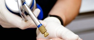
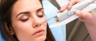
Removing warts with liquid nitrogen
The method of removing warts with liquid nitrogen is now considered the most popular and effective treatment method. The procedure is practically painless, there is no contact with blood, which prevents the spread of infection, and no scars remain. Its essence is that unwanted formations on the skin are frozen during the evaporation of liquid nitrogen and die. Impact doses of cold are a very effective prevention of infection. In addition, this method is the most preferred for removing plantar warts.
Methods of cauterization of erosion
Among the various ways to cauterize erosion, radio wave treatment is considered the most promising, non-traumatic and painless.
However, this method is one of the most expensive, so other types are also in demand and widely used. The best way to cauterize cervical erosion is determined by the attending physician based on factors such as the size of the damage, its location, the presence of dysplasia or tissue degeneration, age, general health of the patient, the woman’s desire to maintain reproductive function and plan a future pregnancy. Since cauterization is often performed under anesthesia, the selection of anesthetic anesthesia is also carried out on an individual basis.
Modern clinical gynecology has a set of methods for physically eliminating the pathological process.
Among them are:
- Diathermocoagulation is a method of treatment with electric current. One of the outdated and most traumatic methods of getting rid of erosion.
- Cryodestruction is a method of getting rid of erosion using nitrogen. This is a more gentle way to remove erosion by freezing pathological cells and their subsequent destruction.
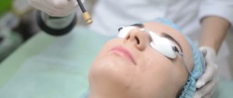
- Laser vaporization is a method of getting rid of pathology using a laser. A fairly painless and effective way to remove erosion, which is highly effective.
- Radio wave coagulation is a method of cauterizing erosion using radio waves. One of the most promising and progressive methods of healing damaged areas.
- The argon plasma ablation method is the elimination of erosion using argon. It is performed using special devices in which argon undergoes ionization with high-frequency currents and precisely affects the eroded area with a plasma beam.
- Electroconization is used to treat severe dysplasia and allows even deep epithelial layers to be removed from atypical cells.
- Ultrasound. Relieving the patient of erosion using ultrasound.
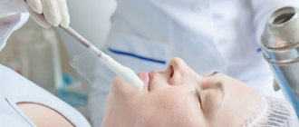
- Chemical or medicinal cauterization. Most often, the drug Solkovagin is used for this purpose, which causes tissue necrosis, the formation of a scab, followed by its replacement by a layer of new epithelium.
Care after removal
After exposure to liquid nitrogen, scars take one to two weeks to heal. It depends on the individual characteristics of the body and the degree of damage. If your doctor prescribes you to take any medications, you should take a course. The bubble that appears on the skin at the site of the session should not be wetted, punctured, and should not be touched at all. Sometimes treatment with potassium permanganate is required for some time after the procedure. The specialist will tell you about this in more detail at your appointment.
Pathomechanism of low temperatures. How does cryotherapy work?
All morphological, biochemical and physiological phenomena accompanying freezing are directly or indirectly a consequence of the formation of ice crystals.
The extent of tissue damage depends on the rate at which the temperature drops, the duration of exposure of the cells to subfreezing temperatures, the lowest temperature reached in the tissue, and the rate of thawing.
Slow cooling of tissues (from -5 to -15 o C) causes the formation of large ice crystals in the extracellular environment. An increase in the concentration of electrolytes in the remaining volume of fluid causes a change in concentration gradients between the extracellular and intracellular environment. This promotes dehydration and cell contraction. These changes can disrupt the phospholipids of cell membranes and disrupt their continuity. The hypertonic environment inside the cell promotes the release of its components beyond the cell membrane. Dehydration of cells also destroys lysosomes.
However, extracellular ice is not the main cause of damage. Rapid freezing causes the simultaneous formation of extracellular and intracellular ice. The number of ice crystals increases as the rate of freezing increases, and their size decreases.
- The presence of ice crystals inside the cell causes damage to mitochondria and the endoplasmic reticulum. Therefore, the primary disorders concern the plasma membranes.
- Secondly, there are changes in proteins and enzymes. DNA synthesis is suppressed.
The higher the freezing rate, the greater the degree of intracellular ice formation and the greater the damage to cells.
The thawing process has a major impact on cell survival. Rapid thawing gives cells a better chance of survival compared to slow thawing. This is due to the process of recrystallization, that is, the combination of small ice crystals into larger ones. Recrystallization is especially enhanced during slow thawing of tissues. The degree of damage increases with increasing crystal volume.
Schematic representation of the hypothesis of two-factor cell damage during freezing and thawing
Therefore, in the case of cryotherapy treatment, the optimal procedure is to quickly freeze the tissue, use a sufficiently long cryo-application time, slow natural thawing (at a rate of no more than 10 °C/min), and possibly repeat the freeze-thaw cycle, which increases the likelihood of cell death.
The direct effect of cryomedrosis is aggravated by the development of vascular changes during and after the procedure. During freezing, blood vessels constrict, which leads to inhibition of blood flow and stagnation of blood in the area exposed to low temperatures.
- Most capillaries are affected immediately.
- In medium-sized vessels (with a cross-section of more than 0.33 mm), circulation stops after a few days.
- In larger vessels, cryosurgery does not cause contraction and they retain their function.
About 30 minutes after freezing, circulation returns, accompanied by a rapid rise in temperature. Dilatation of capillaries and small veins is observed in the presence of blood thickeners and microembolisms. This contributes to inhibition of blood circulation, hypoxia and cell death.
Changes in the ultrastructure of the vessel walls are also observed, causing an increase in their permeability and massive exudate. Bleeding may occur due to the opening of arteriovenous connections in the damaged area. 2-3 hours after freezing, the blood supply decreases again, and this phenomenon intensifies over the next 12 hours.
In the tissue immediately surrounding the freezing zone, an increase in vascular blood flow is observed as the temperature decreases and with greater intensity after thawing, which is manifested by erythema of the surrounding skin.
The described changes in cells and tissues are manifested by a macroscopic image characteristic of cryotherapy:
- During treatment, freezing and changes in the consistency of the area are observed;
- After tissue frostbite, redness appears associated with hyperemia in this area, and then rapidly increasing swelling;
- After a few hours, exudation or a blister appears. The rate at which blisters form depends on the location of the frozen lesion - in the case of a facial lesion, a blister appears within a few minutes, in the case of foot surgery only after a few days.
- The blister lasts about 7-10 days, then a scab forms for 2-3 weeks;
- After the scab falls off, a fresh light pink scar is visible, which after a few months becomes paler and less noticeable.
No anesthesia is used during the procedure. The procedure itself is generally painless; when freezing, the patient may feel a “burning sensation.” Immediately after the procedure, pain may appear during the thawing phase, which quickly passes.
Precautionary measures
Cryotherapy is absolutely safe. When carried out correctly, recovery time takes no more than a day and is not accompanied by restrictions. This distinguishes the method from other hardware cleanings.
It is important that the cosmetologist knows the technique of performing this procedure (they undergo special training for this). Distributing the substance too quickly does not have the desired effect, and the slightest delay can lead to frostbite.
To maintain the result and prevent the appearance of pigmentation, you must:
- use sunscreen;
- choose a soft foam for washing;
- take care of skin hydration.
Side effects
Negative consequences are possible if the procedure protocol is not followed, which is due to the lack of experience of the cosmetologist. If the specialist is sufficiently qualified, complications will not arise.
In rare cases, the following are observed:
- mild redness;
- mild swelling;
- allergy to cold;
- peeling of the skin;
- change in skin color;
- damage to the tissues of the lips or conjunctiva of the eyes.
Contraindications:
- exacerbation of herpes;
- epilepsy;
- heart and vascular diseases;
- open form of tuberculosis;
- the presence of wounds, abrasions, burns on the face;
- individual intolerance to cold.
Signs
So, in order to “see” a wart, you need to know its characteristic signs:
- as a rule, such a formation does not hurt;
- size can vary from several mm to 1 cm;
- the formation may be single or part of a growth (the latter include vulgar warts or genital warts);
- looks like a growth, rises above the skin;
The wart usually rises somewhat above the skin - surface without skin pattern;
- depending on the age of the person, it can be smooth or rough, flaky;
- to the touch is defined as dense, hard;
- the color in young patients does not differ from healthy skin, in older patients it may have a dark color;
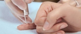
- most often localized on the hands, for example, due to frequent skin trauma.
Warts often appear in the area of the hands
Advantages and disadvantages
| Advantages | Flaws |
|
|
Cryomassage during pregnancy
Pregnant women are allowed to do cryomassage only on the face.
At the same time, given the instability of the hormonal background of women during pregnancy, it is important to ensure that the influence of cold does not affect the tone of the uterus.
If you have a strong desire or need to perform facial cryomassage during pregnancy, be sure to consult your doctor .
We also recommend doing 2 procedures followed by a break to look at the body’s reaction in order to avoid adverse reactions.