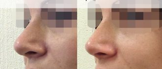
Many of us, trying to get a beautiful and pleasant appearance, resort to various cosmetic surgeries. Rhinoplasty is considered the most common surgical procedure to correct the shape of the nose.
However, the human body tends to provide lost bone strength after surgery, so neoplasms often arise at this site. This causes the development of what is known as a “callus.” Such a neoplasm is quite rare, however, as a result of such a procedure, almost every patient undergoes it. Bone callus has nothing in common with ordinary callus; it does not represent hardened epithelium.
The growth is formed in the event of improper fusion of bones after surgery or nasal fractures, and therefore represents a normal regeneration process.
Why is “bone callus” dangerous?
Such an education does not mean anything serious for life, however, diagnosis and prevention must be carried out in a timely manner. In its absence, serious consequences may occur, including constant severe pain in the patient.
According to statistics, complications occur in 12 percent of victims. In 30 percent of cases, it is necessary to perform a second operation to prevent the proliferation of bone tissue.
When performing rhinoplasty, part of the bone and skin is removed. Operations of this type are always associated with changes in the structure of the nose. During interventions, the human body gives a protective reaction.
Unlike other body systems, bone tissue has a special healing process, which includes 3 successive stages:
- The first is accompanied by the appearance of neoplasms around the site of connective tissue damage;
- At the second stage, the development of bone tissue fibers is noted;
- Then the bone tissue thickens due to fiber calcification. Over time, it completely replaces the soft one.
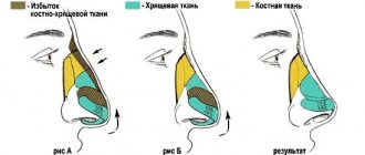
When passing the third stage, a small growth forms right at the site of fusion. If grinding is done during rhinoplasty surgery, then rehabilitation almost always takes place with the appearance of such a callus. The size of the growth always depends on the individual characteristics of the human body, the depth of damage to the bone and surrounding tissues.
Among the main reasons for the development of such a callus are the following factors:
- The presence of individual characteristics of the body, which have a fairly high degree of tissue restoration;
- An equally important factor to ensure a successful operation is the experience of the surgeon. Professional specialists with extensive experience know several secrets that help to carry out the operation in such a way as to prevent bone tissue from growing in the future.
Factors affecting fracture healing
Common factors include: dysfunction of the endocrine glands, pregnancy, vitamin deficiency, acute and chronic infectious diseases, trophic disorders, etc.
Local factors are the leading causes of impaired reparative osteogenesis. They can be divided into three groups (D. V. Ruda, 1976):
1. Errors in treatment: insufficient reposition of fragments and unresolved interpositions of soft tissues between them, unreliable immobilization after reposition and frequent replacement of plaster casts, too extensive bone skeletonization during surgery (blood supply is disrupted), use of inadequate fixators for osteosynthesis (unstable fixation), etc. .
2. Factors associated with the severity of the injury and its complications: multiple and open fractures, massive damage to soft tissues (muscles, blood vessels, nerves), suppuration and osteomyelitis.
3. Reasons that depend on the anatomical and physiological characteristics of the fracture: location, degree of blood supply (fracture of the head or neck of the femur, scaphoid) and others.
Violation of reparative osteogenesis in bone fractures leads to delayed consolidation (fusion) of fragments, nonunion or formation of a false joint (pseudoarthrosis), and sometimes to neoarthrosis (new joint). Delayed consolidation of a fracture refers to cases where bone fusion of fragments has not occurred within the generally accepted normal time frame for a specific location of the fracture.
What does callus consist of?
Callus has a special structure, which appears as a result of the restoration of bone tissue when it is damaged, and is a natural process. We can say that this neoplasm consists of connective tissue growing at the site of bone fusion.
Bone tissue also grows in three stages:
- The appearance of a provisional callus;
- Formation of osteoid tissue;
- Replacement of connective fibers with bone ones.
Almost always, cartilage tissue is formed first, which eventually turns into bone. This process takes about one year.
Non-united fracture
A non-united fracture is one in which, after twice the period required for fusion of a given bone, pain and pathological mobility at the fracture site are clinically detected, and radiographically - a gap between the fragments with the bone-marrow cavities of the fragments still closed (osseous non-union). If there is bone fusion of these cavities with end plates, this indicates a developed false joint (pseudoarthrosis).
So, it is possible to differentiate a non-union fracture from pseudarthrosis clinically by pain at the fracture site, which occurs during movements and loading of the limb, and radiographically by the absence of fusion of the bone marrow cavities.
All consequences of impaired reparative osteogenesis are pathogenetically interrelated and depend on causative factors and the quality of treatment. During the movement of fragments, constant injury occurs to the fresh structures of the callus, including newly formed vessels.
While maintaining the ability of the human body to undergo a reparative process, compensatory changes appear in the area of the fracture in the form of marginal growths, which, to one degree or another, gradually reduce the pathological mobility of the fragments. A hypertrophic or hypervascular callus is formed, in which the processes of bone formation predominate over the processes of bone resorption. Despite the formation of significant fusiform hardening in the area of the fracture, pathological mobility and pain are clinically determined; radiographically, bone fusion between them is not visible. The gap between the fragments is filled with coarse fibrous connective tissue.
Further, the regenerative process with slow fusion can go in two directions, which depends on a number of factors. If the fragments are compressed among themselves, and when they are loaded (physiological muscle contraction, dosed load in a bandage), the acting force coincides with the axis of the damaged segment and goes perpendicular to the fracture line, then the fibrous connective tissue turns into cartilage, and then into bone, i.e. e. secondary bone fusion occurs, although it takes quite a long time to occur.
If the force acts not along the axis of the segment, coincides with or approaches the fracture line, then the bones will not heal, and a hypertrophic false joint will gradually form. Characteristic clinical signs of a pseudarthrosis are pathological mobility and absence of pain at the site of the fracture; radiological signs are the closure of the bone marrow cavities (the presence of locking plates) and the gap between the fragments
The processes of bone tissue resorption predominate over bone formation. The ends of the fragments become thinner and pointed, and the gap between them is wider. Paraosal bone layers disappear. The fragments are connected to each other by connective tissue, which is the least differentiated and does not require a good blood supply. With significant pathological mobility, a gap and typical hypovascular (atrophic) pseudarthrosis are formed between the fragments.
What is the best way to remove a callus after rhinoplasty?
Every person, in case of formation of a callus on the nose, tries to choose the safest methods for its removal.
With hypergrowth of such a formation, the following consequences are possible:
- Ugly hump on the nose;
- Swelling and edema;
- Deformation of the normal shape of the nose.
All of these phenomena not only noticeably worsen the appearance of the face, but also require repeated surgical interventions. Therefore, if the first suspicious symptoms appear, you should visit a plastic surgeon.
Prevention
Preventive measures against callus formation after rhinoplasty:
- Fulfilling doctor's orders during rehabilitation.
- Urgent contact with an observing specialist when the first signs of pathology appear.
- Careful selection of the clinic and surgeon for the operation. Since rhinoplasty is a common procedure and has an affordable price, it is important to analyze all the offers and choose the most proven one with a good reputation, without being tempted by low prices and dubious reviews.
Diagnosis of the disease
The development of callus can be diagnosed using radiography. In the picture it will be visible as a small shell at the site of tissue damage. Rehabilitation is a fairly long and complex process. Its main goal is to stop the growth of bone tissue.
Callus cannot be called a disease, because there is no treatment for it as such. However, there are many different techniques that can improve the patient’s nutrition, reduce the inflammatory process at the site of injury and prevent further growth of bone tissue.
In order to improve the healing of bone tissue, specialists prescribe the following therapy:
- Physiotherapy;
- Surgery to remove bone tissue;
- Treatment with medications.
Removal of callus is considered a radical method and is carried out only in cases where other therapy has not been effective.
Causes of a defect on the nose
The appearance of a callus should be considered as a protective reaction of the bone. After all, surgical intervention involves damage to the internal structure of the nose, and the body begins to regenerate and restore missing elements. The main causes of the defect are considered to be the following:
- tendency to excessive formation of connective tissue with the formation of rough scars,
- unprofessional work of the surgeon.
A similar problem can be detected one year after rhinoplasty. During this time, three successive stages of callus development occur:
- connective tissue appears at the site of damage,
- the formation of thin bone fibers begins,
- calcium salts create a hard build-up.
The size of the formation depends on the scale of the operation performed and the individual characteristics of regeneration.
We recommend reading about closed rhinoplasty. You will learn about the indications and contraindications for the operation, the method of performing it, the recovery period, and the results. And here is more information about the rules of rehabilitation after rhinoplasty.
Features of treatment
The goal of treatment is to eliminate the possible complication. For this, various techniques and methods are used. It is important to use them immediately after surgery. This approach makes it possible to get the desired result faster.
But if there are individual contraindications, effective procedures may be limited. At the first stage, the most important thing is to reduce inflammation at the site of injury using medications.
The tumor can also be removed through surgery. Surgical intervention is a last resort; it is used when other methods have not brought the desired result.
Often this defect after taking medications can manifest itself as the following unpleasant sensations:
- Increase in temperature;
- The appearance of severe pain and redness of the bridge of the nose;
- Difficulty breathing.
Recommendations from specialists are always aimed at effective and safe restoration of damaged tissue. Failure to comply with these rules can lead to serious complications for the patient. Regular visits to the surgeon will help you avoid such complications and undergo rehabilitation faster.
It is important to remember that the body's natural defense system can cause some problems as a result of rhinoplasty. But timely use of physical therapy and medications prevents the need for repeat surgery. As a result of a set of procedures and taking certain medications, the callus quickly resolves.
Taking good care of your body always allows you to maintain a beautiful appearance of your face and the functionality of your nose.
Plastic surgery of the back and tip of the nose without osteotomy
If you want to correct the tip and bridge of your nose, but do not want to make the bridge thinner and the deformation is not pronounced, you will undergo surgery without osteotomy (without surgical dissection of the bone structures). In this case, nose correction will be carried out by influencing the patient’s cartilage.
Correction of the nasal bridge is indicated for patients with a hump, a wide dorsum, or curvature of the dorsum associated with anatomical features or trauma. Often accompanied by correction of the tip of the nose, the wings of the nose, as well as when performing rhinoseptoplasty (correction of the nasal septum).
Often back correction is performed with septoplasty. When the patient experiences difficulty breathing, which is associated, for the most part, not with an aesthetic problem, but with the fact that the curvature of the back is accompanied by problems with the nasal septum. Externally, the nose may “go” a little to the right or left side and cause psychological dissatisfaction with its appearance. In this case, the curved septum does not allow the patient to breathe fully. Lack of oxygen for the body is a serious problem. A person more often suffers from headaches; insufficient oxygen affects all systems of the body, but most importantly, the heart and brain. Correction of the tip of the nose and correction of the wings of the nose can be combined with other manipulations. So, without surgical intervention you can get rid of a whole range of problems.
How is surgery performed in our clinic? When performing rhinoplasty, we use a closed method without osteotomy. We use a unique ultrasound device - a piezo knife (the operation is often called piezorhinoplasty). This method makes it possible to make the operation not only less traumatic, but also a jewelry one. When the surgeon’s work is clear, accurate and coincides with computer modeling up to 95%. This is a very high percentage in rhinoplasty, since the tissues and anatomical features of the patients are different, but the result is always desired and impeccable.
After correction of the nasal bridge, a plaster cast is applied. At the moment, in the usual view, gypsum does not correspond to what it was 10-15 years ago. This is a special flesh-colored pad that fits tightly onto the nose. Wearing a cast is mandatory and lasts from 10 to 14 days; in isolated cases, with complex rhinoplasty and nose reconstruction, the cast can be removed 21-23 days after surgery.
After surgery, it is important to follow all doctor's recommendations. A stable and desired result can only be achieved through joint work between the plastic surgeon and the patient. We are in touch with our patients around the clock, performing the necessary procedures for better healing and reduction of swelling. External swelling goes away within 3-4 weeks, internal swelling can persist for up to several months. The result after surgery should be assessed only after 1 year, but this does not mean that the patient will not see the effect immediately. It is important to understand that tissues and the human body are a complex system and any surgical intervention requires patience. Rhinoplasty is one of the most difficult plastic surgeries. Therefore, trust only professionals and take care of your health.
Taking medications
Drug therapy is considered a fairly common method of treating callus. It is most often used by surgeons as a treatment, as well as for prevention purposes.
In order to prevent the occurrence of hypertrophied callus, it is recommended to use drugs that contain glucocorticoid hormones that can eliminate swelling and ensure rapid bone recovery.
- The most common drug is Diprospan, which is administered by injection. It can increase scarring, relieve swelling and reduce inflammation;
- Another, no less well-known drug is “Kenalog”, the drug is administered intramuscularly, providing an excellent anti-inflammatory effect;
- Also highly popular is a homeopathic drug that has a complex effect on the skin - Traumeel S. The products are used externally or internally; the manufacturer makes them in the form of drops, ointments and tablets.
Physiotherapeutic procedures
Many people prefer to use various procedures that can help restore the nose to its previous shape. Even experienced specialists recognize them as highly effective. With physiotherapy, it is possible to ensure gradual resorption of the callus and also activate its regeneration.
The most common methods of such treatment are the following:
- Using electrophoresis simultaneously with lidase and hydrocortisone preparations. This method can bring a good effect not only at the early stage of callus development, but also with significant compaction;
- Thermotherapy or thermotherapy is also considered effective;
- A good result can be obtained with the help of ultrasound, which is carried out using steroid ointment or phonophoresis;
- It is also possible to use UHF or magnetic therapy.
Everyone understands that it is best to prevent the development of a callus on the nose. It is much easier to carry out thorough prevention with all precautions; in this case, you can avoid such unpleasant consequences, which will then be much more difficult to get rid of.
As preventive measures, experts advise following several rules:
- Contact a surgeon as soon as possible if initial signs or some symptoms of growth development appear;
- Strictly follow all doctor’s recommendations during treatment;
- Choose the right clinic and specialist who will perform rhinoplasty. Such surgical intervention is quite common nowadays, it has a very optimal cost, so you should carefully navigate among the numerous offers in order to choose the most optimal option for yourself.
Recommendations from experienced specialists
To prevent the occurrence of callus, experienced professionals give several tips; by adhering to these rules, you can maintain your appearance as before.
- In the first few days after the procedure, it is recommended to remain in bed; try to comply with this requirement, because this will allow you to get a good result not only regarding your appearance, but also your well-being;
- In the first 2-3 weeks, it is best to stay at home, and you should also try to avoid strenuous physical activity;
- It is not recommended to blow your nose at first; to clean it, it would be much better to use regular cotton swabs;
- Complex strength exercises are not performed; heavy lifting can be carried out no earlier than after 2 months;
- If you wear glasses, you should discard them during rehabilitation. If necessary, they can be worn for a short time;
- Try not to eat foods that are too cold; it is much better to warm everything up;
- For a month you should forget about beaches, saunas, steam baths, and solariums. This will help avoid severe overheating in the sun and under strong steam.
By following all of the above recommendations, a patient who has undergone rhinoplasty will be able to recover quickly and also prevent the occurrence of “callus.”
Surgical treatment of patients with pseudarthrosis
Surgical treatment of patients with pseudarthrosis has been used for a long time, and its methods are being improved as science develops. For pseudarthrosis that formed after a closed fracture, at one time the method of choice was metal osteosynthesis with bone grafting.
After exposing the area, the pseudarthrosis is freed from scars and the bone fragments are refreshed, which, after reposition, are firmly fixed with a metal rod killed by the intramedullary. Then the area of pseudarthrosis is covered with a bone autograft, which is taken from the proximal metaepiphysis of the tibia or iliac wing; allografts (preserved cadaveric bones) or xenografts (bovine bone) are used. The graft is closely fitted with a spongy surface to the exposed layer of the pseudarthrosis area and firmly fixed with wire or bolts. The operation is completed by applying a plaster cast, which immobilizes the limb until the bone heals.
With tight pseudarthrosis without displacement of fragments, good results are achieved using a less traumatic operation - bone grafting with Khakhutov. After exposing the area of pseudarthrosis from the side of the subperiosteal wound, grafts of the same width are cut out from both fragments. Their length in one of the fragments should be 2/s, and in the second - 1/s of the total length of the graft. The grafts are moved so that the longer part covers the pseudarthrosis gap, and the shorter part fills the resulting defect after movement. After surgery, the limb is fixed with a plaster cast until the bone heals.
decortication surgery , has proven its worth . After opening all the soft tissues in the area of pseudarthrosis, thin plates of the cortex are knocked down with a subperiosteal chisel so that they are contained on the periosteum with the adjacent soft tissues. After performing circular decortication, the wound is sutured and a plaster cast is applied.
To stimulate reparative osteogenesis and improve blood supply to the pseudarthrosis area, some surgeons use a chisel to make incisions into the callus and bone to a depth of 2-3 mm in the form of a fir cone. The treatment of patients with infected pseudarthrosis complicated by osteomyelitis and after open fractures was very problematic. Treatment was delayed for many months and even years, since open surgical treatment can be carried out no earlier than 6 months after the healing of the festering wound or closure of the fistula.
To accelerate the healing of infected pseudarthrosis, the Steward-Bogdanov operation, or extrafocal bypass polysynostosis, was used, and for defects of the tibia, the Hahn operation was used - moving the fibula under the tibia.
Ilizarov apparatus into traumatological practice opened a new era, which radically changed the treatment tactics for pseudarthrosis, including those complicated by osteomyelitis and bone defects.
The use of hardware osteosynthesis eliminates deformation, creates stable fixation of the damaged segment, provides movement in adjacent joints, and allows loading the limb. However, with hypovascular pseudarthrosis, the process of bone fusion even in the apparatus remains slow, and therefore it is necessary to additionally apply bone grafting.
Patients with suppurative processes in the area of pseudarthrosis are treated according to the general rules of purulent surgery under conditions of hardware osteosynthesis.
For pseudarthrosis complicated by osteomyelitis , even when there is a fistula, the use of the device and the creation of stable fixation leads to increased regeneration, attenuation of the inflammatory process, closure of the fistula and bone fusion. If there is a formed sequestration, sequestrectomy is performed in the device or before its application. With the help of hardware osteosynthesis, it is possible to shorten the treatment period for patients and achieve bone fusion.
For bone defects, a 4-ring (or more) compression-distraction device is applied, a unipolar, and for large defects, a bipolar osteotomy (compactotomy) is performed in the metaphyseal (cancellous) area of the bone. After the formation of the primary cellular regenerate (7-10 days), the middle bone fragment begins to be lowered towards the defect. Lowering is carried out very slowly, 1 mm per day (in one or two steps of 0.5 mm), bringing the middle rings of the device closer together. As the space in the osteotomy area expands, it is filled with new regenerate and gradually grows.
When the ends of bone fragments approach each other at the site of the former defect, some compression is created to cause necrobiosis and stimulate the local reparative process and fusion of the fragments. For complete bone fusion, the device should be kept in a neutral position for 2.5-4 months. This treatment method allows you to eliminate bone defects over a significant area (15 cm or more).