Patients who want to undergo rhinoplasty spend a long time studying photographs of people who have already undergone the procedure on various resources on the Internet. In the vast majority of cases, they encounter photographs taken after the fact - either chiseled “Hollywood” noses, or outright mistakes of inexperienced or unknown plastic surgeons.
One way or another, minimal attention is paid to when discussing the operation and demonstrating the results . Therefore, the specifics and duration of resolution of nasal swelling after rhinoplasty often surprises, frightens or disappoints those who have not sufficiently studied the information aspect of nose correction surgery.
Rhinoplasty is one of the most difficult and traumatic operations in plastic surgery. During its implementation, various structures of the internal and external nose are damaged - bones, cartilage, mucous membranes, soft tissues and blood vessels.
Swelling after rhinoplasty is an absolutely natural and normal phenomenon that occurs in 100% of cases.
The presence and severity of edema does not depend on the type of correction - open or closed rhinoplasty.
How long does nasal swelling last after rhinoplasty?
The standard period for evaluating the final result of rhinoplasty is one year from the date of its completion.
There are a number of subjective factors that can affect its duration:
- The current age of the patient, which determines the rate of regeneration;
- Individual characteristics of the body (tendency to physiological and pathological edema, metabolic rate, fragility of blood vessels, etc.);
- The degree of density (thickness) and elasticity of the skin on the nose;
- Localization of medical manipulations during rhinoplasty (full rhinoplasty or rhinoplasty of the tip of the nose);
- Patient behavior during rehabilitation.
On average, complete resolution of swelling occurs 9-12 months after rhinoplasty.
Swelling on the inside of the cheek
Anesthesia makes tooth extraction easier. But, if after treatment of a tooth your cheek is swollen, you should find out the origin of the pathology. You may need treatment for your gums. Discomfort appears in many patients after depulpation. Pain and swelling on the inside of the cheek can be observed from 2 hours to 7 days. If discomfort intensifies or occurs 2 days after surgery, you should immediately consult a dentist.
If, after removing the nerve, in addition to painful sensations, the gums become inflamed, purulent discharge appears, and the temperature rises, you should visit a dentist. He will find out why the cheek is swollen and how to remove the gumboil.
Attention! You cannot take painkillers and anti-inflammatory drugs before consulting a doctor; this will complicate the diagnosis, which will not allow you to prescribe adequate treatment.
Swelling after rhinoplasty by month
Swelling after rhinoplasty can be primary and secondary (external and internal).
Primary edema
Primary edema appears during the procedure and disrupts full visualization of the surgical field. For this reason, competent plastic surgeons and anesthesiologists perform local administration of combinations of drugs that enhance the drainage of blood and lymph from the affected tissues. The same manipulation partially neutralizes the primary edema, facilitating the patient’s postoperative adaptation.
From the point of view of sensations, the most difficult thing for the patient is the primary edema. It is much more pronounced than the secondary one, therefore it brings a certain discomfort in the form of a feeling of fullness, pressure and obstruction in the nasal area. Fortunately, the primary swelling is well controlled by a splint (immobilizing bandage) and quickly disappears. After removing the splint, a temporary increase in primary edema is possible, which is neutralized after 1-2 days. Also, due to the retention of swelling by the splint, swelling of neighboring areas of the face is possible - the lower eyelids, cheeks, forehead, and even the chin.
The average duration of primary edema persistence is 7-10 days.
At this stage the patient should:
- Control your sleeping position - sleep only on your back with your head elevated (two pillows or a raised headboard);
- Avoid any physical activity, including housework;
- Limit postures with the head and face lowered forward (when washing, washing hair, etc.);
- Avoid face contact with water;
- Protect your face from sudden temperature changes and especially excessive overheating (sauna, bathhouse, etc.);
- Replace corrective glasses with contact lenses;
- Quit alcohol and cigarettes;
- Stop using skincare and decorative cosmetics.
An important note regarding smoking: nicotine provokes vasospasm, which further increases swelling and slows down regeneration in general. Of course, a plastic surgeon cannot control a patient’s bad habits. But if you want rehabilitation to be easier and faster, try to reduce the number of cigarettes you smoke, or even better, find an alternative in the form of an electronic cigarette with a nicotine-free mixture.
alcohol for the first 2-3 weeks of rehabilitation. In the future, drinking alcoholic beverages is possible, but it will provoke morning swelling of the face and nose throughout the entire recovery period.
During the first week after rhinoplasty, it is advisable to limit social activity to avoid potential injury or respiratory problems.
By 2-3 weeks after rhinoplasty, more than 60% of the primary swelling has disappeared. The face loses its puffy appearance; during this period, most patients resume social activity.
At this stage the patient should:
- Maintain control over the position of the body in sleep - it is no longer necessary to sleep on an elevation, but you still cannot turn on your side or stomach;
- Continue to abstain from strength training, running, heavy lifting and bending;
- Use caution when cleansing your face.
After removing the stitches and plaster
After consultation with the removal of stitches and plaster, consult your doctor about the use of medicinal ointments that stimulate tissue trophism (Traumel, etc.).
The unauthorized use of hot and cold compresses to relieve swelling is strictly prohibited! Such actions can lead to disastrous and painful consequences.
Closer to the second month after rhinoplasty, the primary swelling disappears completely. Only small swellings and induration may remain on the outside of the nose. It is important for the patient to continue to lead a healthy lifestyle, avoid physical inactivity and protect the nose from various injuries.
Secondary edema
Secondary edema is internal swelling that lasts up to a year. It can move, increase and decrease, change the shape of the nose, but it does not bring much discomfort to the patient. Guaranteed resolution of secondary edema occurs by 10-12 months after rhinoplasty, if no complications arise during rehabilitation.
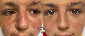
Photo
Visual examples of what swelling looks like after rhinoplasty are shown in photographs of patients.
How long does swelling last after nasal tip rhinoplasty?
There is a noticeable difference between a full rhinoplasty and a tip rhinoplasty. In the first case, bone tissue is necessarily affected, in the second - only cartilage and skin. Due to less trauma, rehabilitation is faster, and swelling may subside by 6 months after surgery. As a rule, patients who have undergone tip rhinoplasty do not notice severe swelling on the face - rather, they are concerned about its hardness and enlargement.
If the plastic surgeon worked with outdated technology, in the early stages of rehabilitation, excessive upturning of the tip of the nose is possible, which then gradually drops. If rhinoplasty was carried out according to a modern scheme (structural rhinoplasty), a rigid supporting structure made of cartilage will not allow the tissues to move and deform, so the tip will remain in the same place, with an ideal nasolabial angle.
Structural rhinoplasty completely eliminates such delayed defects as upturning of the tip of the nose like a “piglet” or its drooping like an “eagle’s beak.”
What does flux look like?
It is of infectious origin, the process occurs against the background of inflammation of the body of the jaw or in the periosteum. Flux is formed not only after dental surgery, but also after furunculosis or tonsillitis. If the cheek and gums are swollen, there is throbbing or mild pain, in advanced stages pus may appear and the temperature may rise. On the upper jaw, the flux covers the lip, cheek, gum and nasolabial area. In addition to the face, the infection often spreads to the neck.
Anti-inflammatory and antibacterial drugs are used for treatment. After using topical medications, you should not eat or drink liquids for 2 hours.
Associated symptoms
In addition to swelling in the first days after surgery, you may be concerned about:
- local pain in the area of the operation;
- redness of the gum tissue;
- temperature rise to subfebrile levels;
- slight secretion of lymph mixed with blood (ichor).
Depending on the volume of the operation, the amount of osteoplastic material introduced and implants installed, the severity of the above symptoms may vary.
The contract for treatment at our Center must indicate all acceptable postoperative conditions. Please read them carefully and the list of possible inconveniences.
Measures to prevent the consequences of sinus lifting in our Center
To avoid situations where swelling turns from a natural reaction into a problem for the patient, we have developed Uniform Quality Standards. We do not work in a rush, we provide personalized dental care, and devote the necessary time to seeing each patient. We provide a lifetime guarantee for bone grafting and implantation operations .
Preoperative preparation
If during the preparation stage obstacles to sinus lifting or bone grafting are identified, they are first eliminated, and only then the day of surgery is set.
Preoperative preparation includes:
- Computer diagnostics A CT scan is performed on a high-precision GALILEOS tomograph in ENT mode to assess the condition of the bone tissue adjacent to the area of the operation of the teeth. On the day of the initial consultation, immediately after the computed tomography, the attending physician, having familiarized himself with the anatomical features, will warn you about possible individual inconveniences and the likelihood of the body’s natural reaction to the intervention.
- Examination by an ENT doctor If diseases of the ENT organs are suspected based on the results of a computed tomography scan, before a sinus lift, an examination is carried out by a regular ENT doctor. If a cyst is detected in the maxillary sinus or inflammatory processes, conservative or surgical treatment is prescribed.
- Sanitation of the oral cavity If any dental pathologies are detected (inflammatory gum diseases, caries, inflammatory formations on the roots of teeth), which are foci of infection, their treatment is mandatory. This reduces the risk of developing postoperative complications of infectious origin. Also, complete sanitation of the oral cavity may include professional teeth cleaning to get rid of plaque and tartar.
Experienced doctors
Bone grafting and sinus lift operations in our Center are performed only by maxillofacial surgeons . These are experienced doctors with continuous work experience of 5 years, deep knowledge of the structure of the maxillofacial system and honed manual skills.
Low-impact technologies
The use of gentle technologies without a hammer or chisel prevents injury to soft and hard tissues and, accordingly, minimizes the occurrence of complications.
- Ultrasonic piezosurgery To work with hard tissues when creating access to the maxillary sinus, we use the NSK VarioSurg ultrasound device. It has a mild softening effect on the bone, without injuring it.
- Using the Lift-Control instrument system A coordinated set of micro-instruments allows you to lift the thin lining of the maxillary sinus as carefully and delicately as possible without damaging it.
- Biocompatible materials and growth stimulants We use only hypoallergenic drugs to increase bone volume in combination with bone morphogenetic protein (BMP). The drugs are certified in Russia and purchased from official representatives.
Maintaining sterility
- Sterilization of instruments is carried out according to the AntiAIDS-AntiHepatitis program. There are 2 separate sterilization rooms, 5 sterilization autoclaving stations .
- All osteoplasty and implantation procedures are performed only in sterile operating rooms. The Center has 3 operating units, which are loaded in a “checkerboard” pattern to allow for sterilization .
Branded rehabilitation
We made sure that each patient was provided with the necessary medications. You will receive a branded package with antibiotics, pain relievers, and wound care products . You won’t have to run to pharmacies after surgery. The package also contains a sheet with postoperative recommendations.
For those who want to go through the rehabilitation period faster, a set of restorative procedures :
- Bioreparation Injections of biostimulants based on hyaluronic acid with amino acids, trace elements and peptides to improve the condition of the skin and reduce swelling.
- PRP plasma lifting Injection of blood plasma into soft tissues to accelerate regeneration processes, improve healing, eliminate swelling and bruises.
- Microcurrent therapy is a physiotherapeutic method of gently applying a weak impulse current to the skin to improve metabolism, improve lymphatic drainage, eliminate swelling, relieve spasms and pain.
24/7 patient support
The post-operative support service operates 24/7. Our specialists will monitor your well-being, remind you to take medications and follow recommendations. This is an additional level of security.
You can ask any questions to the 24-hour medical post support service by calling the numbers listed on the card in the package with medications and recommendations.
Features of rehabilitation after bone block transplantation
When growing bone using the autotransplantation method, donor material is taken from the patient’s body and transplanted to a new location. Therefore, a significant difference from other methods of osteoplasty is that after the operation the patient is left with not one, but two intervention sites. In this case, the patient needs to be as attentive as possible in relation to the oral cavity, carry out hygienic procedures with caution, do not touch the injured areas with a brush and do not pick them with the tongue.