What are nevi
They are a formation consisting of melanocytes. The surface of the mole is smooth and dark in color. There are up to 40 such formations on the body of the average person. Some moles appear on the baby's body immediately after birth. Others are formed during growing up. Nevi can change their appearance and color. Most changes occur during adolescence during puberty. Also, degeneration in a nevus can begin when a person is exposed to direct sunlight for a long time. Ultraviolet radiation in large quantities has a negative effect on the skin. According to statistics, the more time a person spends in the sun, the more moles there are on his body.
The changes concern not only color, but also appearance. An ordinary mole can become flabby and soft, or, on the contrary, develop into a dense and colorless formation. There are a large number of different types of nevi. The main danger of such formations, when exposed to certain factors, is that they can degenerate into malignant ones. Particularly dangerous are complex moles, as well as large formations. They must be constantly monitored by the attending physician in order to prevent a dangerous transformation.
Diagnostics
It is necessary to diagnose melanoma in time and determine the stage of its development in order for the treatment of the disease to be successful.
First, an anamnesis is collected, the doctor learns about changes in the nevus.
Do not take a biopsy or expose the mole to chemical and electrical effects unnecessarily.
To diagnose you need to do:
- Histological examination (a superficial smear is taken if there is bleeding or cracks, provided that the tumor is removed immediately after the diagnostic results);
- Epiluminescent microscopy (examination of a mole using a special apparatus to determine its structure);
- CT (image recording, data processing, comparison with standards).
Types of nevi
There are moles on every person's body. Most often they do not pose any danger. If the quantity exceeds 50 pieces, it is advisable to visit a doctor. Such patients are at risk for developing melanoma. Particularly alarming should be asymmetrical formations with an uneven surface, which have a non-standard color, more than 6 mm in diameter, and began to form on the body already in adulthood.
Congenital
This category includes formations with which a person is immediately born. Congenital nevi vary in shape, color and size. In some cases, the mole may occupy a large part of the child's body. Such neoplasms are cause for concern as they often degenerate into cancerous tumors.
Regular
The category of common moles consists of formations that have a symmetrical shape, uniform color and smooth surface. Most often the color is brown or pinkish. There are no foreign inclusions on the surface. Also, such moles can have a dome shape.
Atypical
Moles with non-standard size and appearance. There are few of them on the body, several times less than ordinary moles. The main danger of such neoplasms is that under certain factors they can degenerate into melanoma. Another feature of such formations is that they have an uneven color, uneven edges, and the nevus itself is asymmetrical. For people with atypical moles, it is important to control their number on the body; the more formations, the higher the risk of melanoma. Constant monitoring by the attending physician and completion of the necessary studies is required.
Blue
Neoplasms are divided into two categories: they can appear on the body already in the womb or develop during a person’s lifetime. They got their name due to their bluish color. But this category also includes nevi, the color of which varies from light gray to black. Most often, formations characteristic of the skin of Mongoloids; in other races they develop in extreme cases.
Miescher's nevi
Formations are brown or flesh-colored. The most common place for moles to appear is in the neck and face. The surface is hard, dome-shaped, but smooth, without foreign inclusions. Hair often grows on the nevus.
Nevi of Unna
In appearance they are similar to the moles from the previous description. Features include their appearance: they resemble raspberries. The color of the new growth is brown.
Meyerson's nevus
It is not difficult to determine the problem around such a mole, a rash with red papules begins to develop and spread. In some patients, eczema in the area is not registered. Another feature of Meyerson nevi predominantly develops in men over the age of 30 years. Women are much less susceptible to this problem.
Nevi halo
They got their name due to the atmospheric phenomenon of the same name. A pale whitish ring begins to appear and develop around the mole. Such education does not last until the end of life, even if nothing is done with it. First the ring changes its color to pink, and then disappears. In some cases, new haloes may appear around the tumor throughout the patient's life.
Spitz nevi
Moles are slightly raised above the skin and have a dome shape. The formation itself is pinkish in color; it is acquired and appears on the skin of patients at a young age. Color may be different. The surface of the nevus is often damaged and begins to bleed. As a result, doctors often confuse such moles with malignant ones, referring the patient for histological examination.
Reed's nevi
The color of the formations varies; they can be black or dark brown (closer to a black tint). Mostly develop in women. The peculiarity of the neoplasm is that it increases in size very quickly, which is why it causes concern among doctors. In fact, such moles are mostly harmless and rarely develop into cancer.
Agminated
They are several moles at once, which are concentrated on a small area of the skin. The peculiarity of formation is that all nevi are not the same. Some may be more, some may be less. Also, among flat formations, dome-shaped ones, etc., may appear.
Only the main types of moles are listed above. In fact, there are a huge number of such neoplasms on the skin. To determine what type of mole a particular mole on the body of a patient seeking medical help belongs to, doctors perform dermatoscopy.
Important features of congenital melanocytic nevi
- Small-sized UMNs are more common and may not appear until age two.
- Small and medium-sized nevi grow more slowly than the child himself and tend to darken and become hairy.
- Large and giant nevi usually occupy part of an anatomical area or all of it, for example an entire limb, neck, part of the back.
- Congenital giant melanocytic nevi transform into melanoma in 6–10% of cases.
Giant VN exceeds 20 cm in size, and such formations are sometimes compared to clothing, called “shirt” or “swimsuit” type. According to statistics, they are rare, affecting 1 in 500,000 newborns.
Giant VNs are only diagnosed at birth. Unfortunately, there is no prenatal screening to detect them, and they are not visualized on ultrasound.
The main medical problem with congenital giant melanocytic nevus is the high risk of developing melanoma, and the tumor can appear anywhere and anytime. What to do in this case? At the moment, the main method is staged excision.
Papillomatous nevus
There are a large number of neoplasms found on the human body. Not all of them are moles. Thus, warts, condylomas, papillomas and other formations may appear. When making a final diagnosis, the whole difficulty lies in the fact that the listed types of neoplasms are often no different in appearance from ordinary nevi. The most problematic is considered to be papillomatous nevus. In appearance, it is no different from ordinary papilloma caused by HPV. But when conducting research, it turns out that this is a bumpy mole of a convex type. The mole is benign, there is no risk of degeneration into cancer. The surface has a flesh-colored, brownish or light brown tint. Dark moles of this type are extremely rare.
A characteristic feature of such moles is the presence of hair on the surface. They mainly appear on the head or neck; they can develop in other parts of the body, but in very extreme cases.
The appearance is not tied to a specific age period. They develop in both teenagers and old people. The mole grows over time, sometimes it increases in size so much that it begins to cause discomfort to the wearer. If the nevus is located on the head, it can be easily damaged when combing the hair. This leads to the onset of inflammatory processes that affect nearby tissues.
The nevus is harmless; its removal is generally carried out only for cosmetic purposes. The greatest discomfort for people is caused by moles on the face, which are visible to everyone around them. Before deciding to undergo a tumor excision procedure, it is important to consult a dermatologist. Only after all the studies have been carried out will the specific type of nevus be established, and the likelihood of the problem degenerating will be assessed. It is almost impossible to independently determine what exactly appears on your body.
Indications for removal may also be permanent damage to the surface of the nevus. Most often, several methods are used to combat education:
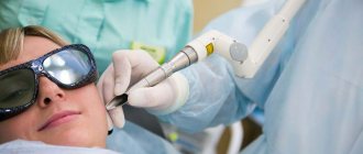
- Laser treatment of the area;
- Freezing and subsequent destruction;
- Radio wave exposure;
- Using electric current;
- Surgical intervention.
If the formation appears on the face, laser therapy is most often chosen.
Methods for removing pigmented nevi
- Small nevi are removed using a laser. Local anesthesia is used here and many small nevi can be removed at once in one visit. A crust appears at the site of removal, which will disappear in seven days. The marks are practically invisible, which is why the laser method is used to remove facial moles.
- For large nevi, surgical excision is used. The operation is performed under local or general anesthesia with suturing.
The decision on the treatment method is made as a result of examining the nevus. In some cases, consultation with an oncologist is required. When surgical excision is used, the tumor is sent for histology to determine whether it is melanoma.
You should never remove moles and birthmarks yourself. This is fraught with infection and other troubles.
Conventionally, pigmented nevi can be divided into four categories according to their size:
- small (up to 1.5 cm)
- medium (up to 10 cm)
- large (up to 20 cm)
- giant (more than 20 cm)
Multiple pigmented nevi are quite rare (5%). This nevus consists of a large lesion and small pigment spots and affects the lower body, limbs, back and chest.
Intradermal nevus
A type of ordinary nevi that have a dome-shaped shape. Such moles rarely appear in childhood, but they are extremely common in the adult population. Up to ten such formations can appear on the body at the same time. Such neoplasms got their name due to the fact that they are located under the upper layer of the epidermis. Often such moles are difficult to notice; they hardly stand out above the surface of the body, and their shade is close to the surrounding tissues.
Nevus can appear in almost any area. Favorite places for neoplasms are the upper part of the arms, eyelids, neck, face, etc. The maximum size of moles is up to 1 centimeter. When they appear in childhood, nevi are practically invisible, but with age they begin to darken and often acquire a convex shape. If the mole persists, then after 70 years it will gradually begin to discolor.
There are several reasons why intradermal nevi can form on the human body:
- Hereditary factors
. If the baby’s parents have more than 50 ordinary moles on their body, then the child’s nevi will also be multiple in nature. - Exposure to sunlight
. Ultraviolet radiation negatively affects the skin, causing damage. Moles often appear in place. People with too light skin are at risk. - Decreased immunity
. Most often, nevi begin to form against the background of reduced immunity, including after taking drugs that suppress the immune system.
Even ordinary moles that have a symmetrical appearance and do not increase in size are important to constantly monitor. It is better to have a checkup with a dermatologist at least once a year, this way you will be able to identify the problem at an early stage of its occurrence. It is worth planning a visit to a specialist outside of the schedule if the mole begins to grow, changes its shape, acquires a different shade, or begins to bleed.
A dermatologist may prescribe a procedure for excision of a mole if it causes cosmetic discomfort to the wearer, if there is a risk of the neoplasm degenerating into melanoma, if the surface of the nevus is constantly damaged during the day.
Congenital melanocytic nevus is a benign proliferation of cutaneous melanocytes that is clinically observed at birth or appears during the first weeks of life. Congenital nevi vary in size, macroscopic appearance and histological characteristics [1] and appear in approximately 1% of newborns. Most nevi are small in size, but there are large congenital nevi (LCN) and giant congenital nevi (GCN), which have a significant area and sometimes extend to an entire segment. GVNs are relatively uncommon congenital malformations that, when examined macroscopically, are usually characterized by intense pigmentation and can often have variable hair patterns [2].
In histological examination, GVN are characterized by an abundance of small and medium-sized “nevus cells” (melanocytes), with a relatively small amount of weakly oxyphilic cytoplasm, moderately basophilic nuclei of predominantly irregular ovoid shape. In the upper layers of the dermis (the area of the papillary layer), nevus cells often form clearly demarcated, irregularly rounded nested clusters of different sizes and contain a finely clumped brown pigment (melanin) in the cytoplasm. In the deep layers of the dermis, nevus cells of the HVN extend up to the border with the hypodermis and can penetrate into it, as through fibrous septa separating the cells of the subcutaneous fatty tissue, and appear directly in its tissue. An increase in the number of macrophages in nevus tissue and their increased activity, expressed, in particular, in the increased absorption of melanin pigment by macrophages, is reported [2].
Congenital giant pigmented nevi are often a significant cosmetic problem and a significant psychological traumatic factor for the child. The particular significance of giant pigmented nevi is also due to the fact that they are characterized by the potential for transformation into a malignant neoplasm - melanoma [2-4]. There is an opinion that malignant melanoma, developing from other types of nevi, usually arises in places of the epidermal-dermal junction (in the superficial layers of the skin), while melanoma from the GVN originates mainly in the deep layers of the dermis [2-4], which is obvious , is a condition that preserves the possibility of malignancy of the remnants of the GVN when it is incompletely removed. The reasons for the emergence and development of GVN are discussed, although there is a fairly well-established opinion according to which GVN develop from primitive cells of neuroectodermal origin (from neural crest cells) that are delayed during migration and subsequently proliferate [5]. There is also evidence that these tumors differ in a variety of morphological manifestations and clinical behavior [6]. Due to the fact that most congenital giant pigmented nevi undergo early surgical and therapeutic treatment for cosmetic reasons and for prophylactic purposes, the true incidence of their malignancy is difficult to determine [2].
There is also no consensus on the need to remove nevi and an unambiguous view on the choice of treatment method. Currently, several basic methods for eliminating WMH are described in the literature and used in practice: staged excision with plastic surgery using a free split or full-thickness autograft, the use of flaps obtained by tissue dermatosis, dermabrasion, curettage and laser [7]. A number of authors [8–10] report on surgical treatment of GVN using microsurgical techniques.
The purpose of the study is to study the clinical and histological features of nevus tissue, which are important for the choice of surgical intervention technique, immediate and long-term results.
Material and methods
During 2008–2010, we examined and operated on 10 patients with VVN and GVN of different localization. The age of the children ranged from 9 months to 7 years. When formulating the diagnosis, we used the classification proposed by foreign authors, according to which a nevus constituting less than 1% of the body surface area on the face and less than 2% on the body is considered BVN, and more than 1 and 2%, respectively, is considered to be GVN [1, 8].
All admitted children were registered with an oncologist and had a conclusion that there were no contraindications to surgical treatment. As part of the preoperative examination, all children were examined by a neurologist and a neurosurgeon. According to indications, magnetic resonance imaging of the affected area was performed. The examination did not reveal any neurological abnormalities or concomitant developmental anomalies.
Surgical treatment included staged excision of the nevus with replacement of the defect with local tissues in the form of rotational flaps, thick split or full-thickness skin autograft. The choice of donor site depended on the location of the surgical wound. For facial transplantation, full-thickness autografts from the inner surface of the shoulder were used. The donor wound was closed with a split autograft from the anterior inner surface of the upper third of the thigh. For plastic surgery of defects of other localization, excess skin obtained by the method of expander dermatension and split autografts from the anterior inner surface of the thigh were used. Removed skin fragments from GVN and BPN were studied using morphological research methods: macroscopic description, light microscopy, morphometric calculations of certain indicators and, in some cases, using immunohistochemical (IHC) reactions.
results
The main clinical symptoms of nevi (except for the presence of the GVN itself with its macroscopic characteristics) were a feeling of itching in the affected area, a periodic burning sensation and peeling. Itching most often bothered patients in the area of nevi with hairline, was obsessive in nature and was accompanied by scratching and trauma. The most pronounced hair growth was observed when the nevus was localized in the lumbosacral region (Fig. 1).
Figure 1. GVN in the lumbosacral region. In 2 patients, new small pigment spots were noted on different parts of the body within 1-1.5 years after birth, which may indicate the continued mitotic (proliferative) activity of melanocytes [11].
In case of multiple nevi, surgical intervention was primarily performed on the GVN and BVN, located in areas of the body that experience increased stress and are most susceptible to trauma (hands, feet, Achilles tendon area; Fig. 2).
Figure 2. BVN located on the thighs, legs, Achilles tendon area, and feet. Priority removal was also given to the GVN and BVN, which were located in open areas of the body most susceptible to solar insolation (face, cranial vault, upper limbs). Then the largest nevi of another location or nevi that bothered the children were removed. Since warty nevi were the most frequently traumatized, they, in our opinion, must be removed regardless of size and location (Fig. 3).
Figure 3. Warty nevi.
When choosing a method for closing a wound defect, the age and constitutional characteristics of the child, the thickness of the skin, and the severity of the subcutaneous fat layer were taken into account. In children under 3 years of age and with an asthenic constitution, large-volume expanders were placed on the cranial vault and chest area to avoid deformation of bone structures, the development of trophic complications over the domes of the expanders, and limiting respiratory excursions of the chest.
At the first stage of surgical treatment, we tried to maximize the use of local tissue reserves to close the defect formed after excision of the nevus area.
The next stage was planned depending on the localization. If the nevus was located in an area suitable for placing an expander, expander dermatension was performed, followed by using the resulting supply of integumentary tissue to close the defect after removal of the nevus. In other cases, free full-thickness or split-thickness autografts were transplanted.
Already during the surgical intervention, significant differences in the appearance of intact skin and nevus tissue were noticed. The latter was characterized by a loose consistency and differed from leather in its reduced strength and reduced elasticity. The subcutaneous fatty tissue also differed from the usual: it was more yellow in color, easily fragmented and was quite flabby.
During macroscopic examination of the surgical material, in almost all cases, changes were observed that were characteristic of nevi in general and of GVN and BVN in particular. The skin fragments were of different sizes and shapes. The surface of the removed skin flaps had a gray-brown, brown or dark brown color. Already at the stages of visual assessment of the material, it was noted that nevi (small and relatively medium-sized and even some large ones) were removed within intact tissues: around the nevi there was a clearly visible, narrow or narrow grayish rim of little changed skin. Fragments of especially large nevi and GVN in a number of cases were removed within the altered tissues. Some of the skin flaps had a slightly raised outer surface that was quite smooth, in some cases it appeared finely tuberous, sometimes with small papillary formations. In some cases, the surface of the nevus was rough and dirty gray in color. On the section, most often the superficial layers of the dermis and the epidermis region (total thickness from 0.03 cm to 0.08-0.1 cm) had a gray-brown color, the deeper areas of the dermis were whitish. The remains of yellowish subcutaneous fat retained their cellularity.
During microscopic examination, almost the same type of changes were observed in all preparations from fragments of nevi with subcutaneous fatty tissue.
The epidermis retained a fairly well-defined stratification; in some places, moderately pronounced hyperkeratosis was noted (Fig. 4, c),
Figure 4. Giant pigmented nevus (hematoxylin and eosin staining). a — “nest” accumulations in the papillary layer of the dermis of melanocytes containing brown lumpy pigment (×100); b - continuous extensive “fields” of nevus cells in the reticular layer of the dermis (×50 and ×200); c — moderately severe hyperkeratosis (×100); d — small papillomatous structures (×50). Small papillomatous structures were often encountered (Fig. 4, d).
Melanocytes of the basal layer of the epidermis and Langerhans cells were present in the usual numbers and in the usual proportion with the cells of the stratified squamous keratinizing epithelium. During an IHC study, a pronounced positive expression of antibodies to S-100 antigens was observed in these cells (Fig. 5, d),
Figure 5. Giant pigmented nevus: IHC study. Positive expression of the S-100 antigen in nevus cells of the dermis (a, b), in the connective tissue layers in the subcutaneous tissue (c) and in Langerhans cells of the epidermis (d). CD1a (Fig. 6, b),
Figure 6. Giant pigmented nevus: IHC study. Negative expression of CD1a antigen in nevus cells of the dermis (a) and its positive expression in Langerhans cells of the epidermis (b); brown clumpy pigment (melanin) in nested clusters of nevus cells in the papillary dermis (c). and langerin (Fig. 7, b),
Figure 7. Giant pigmented nevus: IHC study. Negative expression of Langerin antigen in nevus cells of the dermis (a) and its positive expression in Langerhans cells of the epidermis (b); brown clumpy pigment (melanin) in nevus cells in the papillary dermis (c). and in melanocytes of the basal layer - also Melan A (Fig. 8, b)
Figure 8. Giant pigmented nevus: IHC study. Positive expression of the Melan A antigen in nevus cells (a) and in part of the epithelial cells of the basal layer of the epidermis (b). and MITF (Fig. 9, b).
Figure 9. Giant pigmented nevus: IHC study. Positive expression of the MITF antigen in nevus cells (a) and in part of the epithelial cells of the basal layer of the epidermis (b). Quite thin acanthotic cords extended from the epidermal layer into the dermis.
The papillary layer of the dermis was often noticeably thinned and smoothed, its papillae preserved only in some places. The reticular layer of the dermis appeared significantly thickened. In both the papillary and reticular layers of the dermis, an abundance of nevus cells—melanocytes—was observed. Melanocytes (Fig. 4, b) are homogeneous, rather large cells, with moderately expressed light eosinophilic cytoplasm and relatively large, light, irregularly round, irregularly oval or “bean-shaped” nuclei. Some nuclei had small single nucleoli.
In the papillary layer, nevus cells form clearly defined “nest” clusters of varying sizes. Most of the cells in them contain a lumpy brown pigment in the cytoplasm - melanin (Fig. 4, a). In the reticular layer of the dermis, the cells are located compactly, in continuous extensive “fields” (Fig. 4, b), and only in some places (mainly in the marginal zones of the nevus) they form numerous clearly defined clusters, separated by fibrous layers. The ratio of nevus cells and collagen fibers of the stroma is sharply shifted towards nevus cells. In the deep layers of the dermis, down to the subcutaneous tissue, nevus cells also form continuous “fields”, but are located less compactly (Fig. 10, a):
Figure 10. Giant pigmented nevus (hematoxylin and eosin staining). The abundance of nevus cells in the deep layers of the dermis, directly adjacent to the subcutaneous tissue, and in the fibrous layers of adipose tissue (a; ×100) and small accumulations of melanocytes in the cells of subcutaneous fat (b; ×100 and ×200). When morphometrically calculating the proportion of stroma in the upper 2/3 of the dermis, it was revealed that the connective tissue stroma on average occupied only 5.4% of the section area, while the average proportion of stroma in the lower 1/3 of the dermis reached 44.4%.
Often, during histological examination of GVN and large nevi, a vertical distribution of melanocytes was observed in depth down to the subcutaneous fatty tissue and along the fibrous layers (Fig. 10, a), extending from the reticular layer of the dermis, further into the subcutaneous tissue. Individual nevus cells were also found directly between lipocytes in the cells of the subcutaneous tissue (Fig. 10, b). The prevalence of melanocytes in small and small nevi rarely reached the hypodermis and even the deep layers of the dermis.
During an IHC study, a pronounced positive expression of antigens S-100 (Fig. 5, a-c), Melan A (Fig. 8, a) and MITF (Fig. 9, a) was observed in all nevus cells, which indicated their melanocytic nature.
The morphological characteristics of some melanocytes (an oval or bean-shaped nucleus, a longitudinal cleft or feature that makes the nuclei of some cells look like coffee beans) suggested that at least some of the cells in the nevus tissue would express antigens characteristic of Langerhans-type cells. However, the reaction of antibodies to CD1a (Fig. 6, a) and langerin (Fig. 7, a) antigens in melanocytes was almost completely negative. At the same time, pronounced expression of these antigens was observed in Langerhans cells of the epidermis (Fig. 6b and 7b, respectively).
During an IHC study, only single nevus cells with positive expression of the Ki-67 antigen were found among the melanocytes of the GVN (Fig. 11, a),
Figure 11. Giant pigmented nevus: IHC study. Positive expression of the Ki-67 antigen in single nevus cells of the dermis (a), its positive expression in some epidermal cells (b) and in epithelial cells of the hair follicles (c). which, along with the virtual absence of mitotic figures in melanocytes, confirms their low proliferative activity in the studied material. More often, positive expression of the Ki-67 antigen was observed in the cells of the stratified squamous epithelium of the epidermis (Fig. 11, b) and in the epithelial cells of the hair follicles (Fig. 11, c).
The cellular subcutaneous fatty tissue removed along with the nevus was formed from highly differentiated mature lipocytes. Its cells are separated by fibrous cords (septa) extending from the dermis, in which numerous nevus cells were often detected. Also, nevus cells could be seen directly between lipocytes in fiber cells (Fig. 10, b).
Thus, histologically, the nevus tissue had significant differences from normal skin. In all sections, weak expression of stromal elements and the absence of the usual appearance of collagen and elastic fibers were noted. In a number of cases, nevus cells spread along the septa extending from the dermis into the subcutaneous fatty tissue, which, according to K. Kishi et al. [7], causes an increased risk of repigmentation (i.e., continued growth of remaining tumor fragments and, accordingly, their possible malignancy). The histological data we obtained indicate that the spread of nevus cells into the subcutaneous tissue is mainly characteristic of BVN and GVN, while medium and small nevi often do not extend beyond the dermis. The identified trend is consistent with the observations of a group of foreign authors [7]. We observed one case of repigmentation after staged removal of the facial vein and replacement of the defect with a thick split autograft. To prevent this complication, it is recommended to perform curettage of subcutaneous fat tissue in order to remove remaining nevus cells; however, for obvious reasons, this manipulation is not recommended on the face, so we consider the maximum use of local tissue on the face to be optimal.
Signs of cellular atypia characteristic of a malignant tumor were not found in any case. According to visual control carried out intraoperatively, nevi were removed along the periphery and in depth within intact tissues. However, in some cases, the spread of nevus cells along the dermal septa into the subcutaneous fatty tissue (hypodermis), revealed during histological examination, cast doubt on the radicality of excision of the nevus tissue.
The described clinical and histological features of the pathologically altered dermis, in particular a significantly reduced content of stromal elements (collagen fibers), indicate an increased risk of developing failure of the intradermal surgical suture when performing staged excision of a large and/or giant nevus, which dictates the need for careful suturing of the surgical wound and ensuring rest of the operated area in the postoperative period.
When the nevus is localized in the middle and lower parts of the face, its staged excision with the application of surgical sutures between the changed and intact skin often leads to failure of the suture or to a gradual expansion of the scar line within 6 months after surgery (Fig. 12, a, b).
Figure 12. Failure of the suture (a) and gradual expansion of the scar line (b) within 6 months after staged excision of the nevus with the application of surgical sutures between the changed and intact skin (localization of the nevus in the middle and lower parts of the face). This is due to gravitational ptosis of the lower edge of the suture and, in some cases, the inability to provide rest to the operated area. The cosmetic effect can be improved with the help of a compression mask, however, as practice shows, the child does not always behave adequately to medical recommendations. The use of combined skin grafting (thick split-thickness or full-thickness autograft) in the middle and lower parts of the face is also associated with an increased risk of developing wide scars due to differences in the thickness and elastic properties of local and grafted tissues.
Excision of a large area of nevus followed by simple suturing of a wound on the face is impossible due to the risk of developing asymmetry and deformation. With this localization of the nevus, it is necessary to strive for the most complete excision of the nevus using large rotational flaps from the zygomatic, cheek, parotid and submandibular areas with sutures between healthy tissues, including using an intradermal suture.
The use of an intradermal suture allows one to achieve good cosmetic results provided that the nevus is removed within intact tissues. If a suture is placed in a functionally active area, we recommend immobilizing the segment with a plastic splint until reliable consolidation of the suture edges occurs. In case of multiple nevi, in case of their radical removal, it is necessary to strive for the maximum use of intradermal technique when suturing the wound.
We used excision of the BVN and GVN followed by combined skin plasty when the nevus was localized in the upper parts of the face - the frontal and paraorbital regions (Fig. 13, a-d).
Figure 13. Excision of the BVN and GVN (localization of the nevus in the upper parts of the face - in the frontal and paraorbital region) followed by combined skin grafting: good immediate and long-term cosmetic results. a, c - before surgery; b, d — after surgery. Good immediate and long-term cosmetic results were obtained.
Thus, when performing surgery, we recommend:
1) priority removal of nevi in functionally active areas;
2) maximum use of local tissues on the face in the form of rotational flaps with intradermal sutures;
3) removal of BVN and GVN within normal skin around the circumference and up to the fascial layer in depth;
4) immobilization of the limbs in young patients when applying intradermal sutures on the limbs and during staged excision of the nevus, when the remaining nevus tissue is included in the suture.
All operated children were observed by us for 2 years. Functional and aesthetic results remained stable.
Epidermal nevus
As the name implies, this category includes moles that are formed from the upper cells of the epidermis. Mostly, neoplasms appear on the child’s body after birth; the vast majority of such nevi also appear in the first year of the baby’s life. A distinctive feature of the neoplasm is that it remains unchanged in shape and size throughout a person’s life.
Epidermal nevi are divided into several types depending on the cells that make up them. The most unpredictable are considered to be formations that include not only keratinocytes, but also sweat gland cells. The formations have a yellow-orange tint; in 25% of cases they degenerate into malignant tumors.
Non-pigmented moles
Most species are characterized by a color ranging from light pink to black. There are a few exceptions to the rule. So, sometimes a nevus may not have pigment. Such moles are easy to identify; they appear as a whitish or lightish spot with unclear boundaries and an irregular shape. Moles without pigment can only appear on the body of Europeans.
Moles without pigment are characterized by a small size of up to 2 centimeters. Basically, only the cosmetic side of the issue becomes an indication for removal. If a person often sunbathes, the nevus remains light in color and, as a result, stands out on the skin and attracts attention.
Treatment method
Today, you can get rid of pigmented nevi in various ways. Medicine offers several options:
- surgical intervention: the formation is excised using a scalpel, while the doctor captures healthy areas of skin and tissue. Sutures are placed at the intervention site, and after they are removed, unfortunately, a scar remains on the skin;
- laser: using a directed laser beam, the formation is removed in a few minutes, leaving a barely noticeable white spot on the skin;
- cryodestruction: the technique is based on exposing the pigment spot to critically low temperatures. There are no cicatricial formations or unaesthetic scars left on the skin;
- radio knife: a minimally invasive, painless method of removal, after which there will be no scars on the skin.
After any of the listed options for removing formations, histology of the removed tissue is required to determine their benignity/malignancy.
Borderline nevus
The formations got their name due to the fact that they often develop into malignant formations. It is important to constantly monitor such moles and contact a dermatologist if any changes are detected.
Such moles are hidden under the stratum corneum of the skin; they have a uniform brown color and clear boundaries. The following factors should cause concern in the patient and provoke a visit to a dermatologist:
- the mole begins to increase in size;
- changes its color, becomes darker or lighter;
- pigment spots of various types began to appear around the adjacent area;
- the mole has become dense, standing out above the surface of the skin;
- Soreness appeared in the mole, the surface was very itchy.
Prevention of melanoma
To avoid skin cancer, you should follow some rules:
- do not be in the open sun, especially from 10 to 16-17 hours;
- do not visit the solarium;
- wear wide-brimmed hats in the summer heat and, if possible, wear clothes made of light materials that cover the places where there are nevi;
- Do not injure the area of the pigment spot, do not irritate it with chemicals.
Do not forget: the use of ointments, creams and sprays against UV radiation does not provide one hundred percent protection against the appearance of melanoma! Take care of yourself, and if a problem occurs, contact your doctor immediately.
Therapist of the highest category, cardiologist, gastroenterologist. Menzhevitskaya Tatyana Ivanovna
What to do with huge nevi
The overwhelming majority of nevi are small formations on the skin with a diameter of several millimeters, which are practically invisible to the naked eye. Exceptions are formations that exceed 20 centimeters in diameter. They are classified as huge. The mole can cover most of the arm, leg, back, etc. Such moles can appear on the body of representatives of any race. They develop in almost 2% of the population of our planet. The main danger of large nevi is that they often degenerate into malignant ones. Formations that appear in the area of the spinal column are considered extremely unstable. Moles that continue to rapidly increase in size and also change their color should cause concern.
The most unpleasant huge nevi on the body of newborn children. As the child grows over time, it seems to parents that the mole is decreasing in size, but in fact it continues to grow, almost not keeping up with the whole body.
In 30% of cases, such nevi become melanoma. They should be under the supervision of a doctor. Rebirth can occur at absolutely any age. Removal of giant nevi is indicated. The most commonly used method is surgical excision. If the mole is located in a hard-to-reach place, the patient may be offered alternative treatment methods. Removal is also possible using laser therapy.
How are nevi treated in the Rassvet blade?
The doctor will examine the patient's skin completely to eliminate the risk of missing melanoma or other skin cancer. Suspicious formations are further studied using a device that allows you to examine the skin with 10 times magnification - a dermatoscope. The patient's chart describes nevi that require observation. The doctor will suggest surgical removal of suspicious nevi, followed by histological examination of the removed material (sent to an expert laboratory). Removal of nevi for aesthetic reasons can be performed at the request of the patient.
Recommendations of a dermatologist for patients with nevi:
- avoid excessive ultraviolet irradiation of the skin (tanning, sunbathing, solariums, ultraviolet drying of manicure and pedicure coatings); when staying outdoors for a long time, use a broad-spectrum photoprotective cream with SPF 30 or higher, clothing that covers the skin, a hat and glasses with an ultraviolet filter;
- try not to injure nevi when shaving, combing, changing clothes;
- undergo an annual preventive examination of nevi with a doctor;
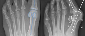
- independently examine nevi according to the ABCDE rule;
- if there are a large number of nevi (50-100 or more), especially in combination with the presence of risk factors (sunburn, visits to a solarium, melanoma and skin cancer previously identified or in close relatives, the presence of atypical nevi), undergo a digital skin mapping procedure.
Author:
Kuzmina Tatyana Sergeevna dermatologist, Ph.D.
Nevi on the head
Such formations are most often removed precisely for cosmetic reasons. If a mole is located on the surface of the body, no matter what size it is, it can almost always be hidden under clothing. Nevi on the head cannot be hidden. Some patients believe that a mole emphasizes their individuality, others are sure that such a formation only spoils their beauty. As a result, patients often seek removal of even those nevi that will never become malignant.
Often, not only moles on the head are removed, but also tumors on the neck. The main danger of such moles is that they often rub against the surface of clothing and are eventually damaged, which increases the risk of degeneration. On the head, nevi also appear in the hair area. They are practically invisible to the naked eye and are often felt by the patient during hair washing and other procedures. The main danger of neoplasms is that they can be easily damaged during banal combing of hair. Nevi, which include cells of the sebaceous glands, also often appear on the scalp in the hairline area. They are characterized by the shape of a wart with no hair on the surface, as well as an irregular shape. Human papilloma viruses, as well as hereditary factors, can cause the appearance.
Most moles on the head appear at birth. The formation is examined and research is done to make a final diagnosis. In childhood, neoplasms are rarely removed, since they are not subject to degeneration during this period. It occurs most often during puberty. The removal method is selected depending on the size of the formation. If the nevus is small, you can get rid of it with laser therapy; to combat large formations, surgery will be required.
It is important to remember that at the moment there is no magic pill that could remove all dangerous moles from human skin that can become malignant. It is important to monitor the condition of nevi and pay attention to any changes.
Moles are successfully removed. It all depends on the size, qualifications of the medical specialist, as well as the location of the nevus.
The child's mole is growing. What to do?
Acquired melanocytic nevi (AMN) are benign tumors that arise from melanocytes that have migrated into the skin. They usually appear after six months of life, and reach their maximum size and number at a young age. Subsequently, they may regress or disappear altogether.
The localization of acquired formations is varied. They can be on the skin of the scalp, palms, feet, and also come from the nail matrix, creating difficulties for diagnosis and observation.
Factors influencing the appearance of nevi:
- genetic predisposition;
- level of ultraviolet radiation in childhood;
- phenotypic features of the child’s skin (fair skin, eyes, blond or red hair).
The classification of acquired nevi is varied and includes typical and atypical forms. They are also classified based on the location of the melanocytes.
PMNs are characterized by a round or oval shape and have clear boundaries. Normally they are symmetrical in color, structure and shape.
Most PN are benign and do not require any intervention, but only require lifelong monitoring.
The frequency of malignant transformations in PMN is low, since melanomas most often develop on clean skin, i.e. beyond previous melanocytic nevi. Therefore, their removal for preventive purposes is impractical.
It is worth noting that the tactics for managing patients with melanocytic formations in childhood can be implemented in three ways: surgical excision of the element, dynamic observation of the formation, and zero-intervention tactics, when further observation or surgical intervention is not required. The doctor makes a decision based on an analysis of all factors characterizing the formation: the child’s age, morphology, location, size and melanoma danger of the element.
An important diagnostic point when examining skin formations is to perform dermatoscopy.
Modern technologies have allowed doctors and patients to “monitor” the formation using its mapping. This is a procedure for fixing moles, which makes it possible to evaluate the dynamics of changes in structures, sizes and the appearance of new formations.