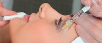
Non-surgical rejuvenation of the neck and décolleté (BEFORE and AFTER photos). Contour plastic surgery, biorevitalization of the décolleté area. Vector lifting: is it possible to lift the décolleté area without surgery?
Plastic surgeon, Candidate of Medical Sciences E. S. Kudinova
When a surgeon talks about rejuvenation, they usually talk about surgery. This is not because “plastic surgeons can’t do without a scalpel.” It’s just that in women 45 years of age or older, surgery can obviously give a good result for a very long time. Any cosmetic procedure (almost always!) cannot be compared with surgical correction neither in terms of effectiveness, nor in long-term effect, nor even in cost (try to calculate the total cost of a non-surgical alternative - injections, threads, laser and other anti-aging procedures that will be required within 15- you years old!). But the rejuvenating effect of the operation lasts all these 15-20 years!
Why NOW do I want to talk not about operations, but exclusively about NON-SURGICAL LIFT?
The fact is that we do not have surgical methods for rejuvenating the décolleté area. So there is simply no alternative to non-surgical methods for correcting this area!
Introduction
The use of contour injection plastic surgery using fillers of various compositions is today one of the most common minimally invasive interventions in aesthetic medicine, second only to the use of botulinum toxin. The main material currently used for these procedures are hyaluronic acid (HA) fillers; their use has been growing steadily over the past 20 years. According to ASPS (American Society of Plastic Surgeons) statistics, the frequency of use of HA fillers increased by more than 2.5 times from 2000 to 2014. The authors’ personal observations from 1998 to 2018 demonstrate even more impressive trends: over 20 years, the number of procedures using HA fillers increased from 193 in 1998 to 1027 in 2022, which represents a more than fivefold increase in the number of contour injection procedures plastics using HA gels (Fig. 1).
Rice. 1. Number of injection contouring procedures using various products.
The history of the use of substances for contour injection facial plastic surgery goes back about 130 years. At the dawn of modern aesthetic medicine, paraffin, fat grafts and silicone were used for procedures. However, the safety and effectiveness of such interventions have not been satisfactory. In the second half of the last century, the first fillers based on bovine collagen (Zyderm, Zyplast) were developed. In the 90s, drugs based on fibrillated Teflon and fibrin were also released. In addition, other collagen derivatives, including human collagen (isolated from the placenta), as well as biopolymers and combined materials, have been marketed at various times. Fillers based on hydroxyapatite and polymers of lactic acid (Poly-L-Lactic Acid - PLLA) have been developed and are used. However, the leading place in the market of drugs for contour injection plastic surgery today is occupied by recombinant HA substances of various modifications [1].
A number of factors have contributed to the spread of HA-based fillers for facial contouring injections. These materials combine a number of advantages that are critical for injection contouring. First, synthetic HA has relatively low immunogenicity. Secondly, despite the limited scientific data on the ratio of the number of adverse events when using different drugs, according to expert estimates, complications with the use of HA fillers are less common. For this reason, the US market is currently dominated by FDA-approved (Food and Drug Administration) drugs based on synthetic HA, the safety of which has been proven in numerous clinical studies [2–4]. Third, HA-based fillers provide significant clinical benefits, such as a long-lasting effect of up to 24 months. depending on the drug, as well as positive results of correction from the patient’s point of view [4]. Despite the faster elimination of HA from the body, fillers based on it provide the most optimal combination of safety and effectiveness for practical use today [5–12].
At the same time, the increased use of HA fillers is associated with an increase in procedure-related adverse events. Thus, in clinical practice in 1998, the authors recorded 3 cases of complications of contour injection plastic surgery with the use of HA, while in 2022 this value was 26, which is an 8-fold increase in the number of adverse events (Fig. 2).
Rice. 2. The number of complications of contour injection plastic surgery when using various fillers. The contribution of various fillers to the overall structure of adverse events associated with their use is presented in Fig. 3.
Rice. 3. The share of various fillers in the structure of complications. For this reason, we decided to conduct a review of modern ideas about the complications associated with the use of HA in facial injection contouring, as well as the tactics of managing patients with various adverse reactions. The article provides information from international expert consensus on complications associated with the use of HA fillers, as well as data from many years of clinical experience of the authors of this article.
Benefits of contouring
- Safety. Hyaluronic acid gels are biologically inert preparations of non-animal origin that do not cause allergic reactions and are absolutely compatible with the tissues of the human body;
- The result of aesthetic correction made with gel fillers is visible immediately;
- The rejuvenation effect after contouring is as natural as possible;
- The procedure does not require anesthesia, unlike plastic surgery;
- No scars or marks on the skin;
- A large amount of work can be done in one contouring procedure - for example, increasing the volume of the lips, eliminating deep wrinkles and emphasizing the cheekbones;
- There is no rehabilitation period. Slight swelling is possible for 2-3 days;
- The result lasts up to 12-18 months, and the use of some drugs helps to extend it up to three to four years.
Classification of adverse events
The accumulated practice of using HA-based fillers has led to the emergence of a large number of expert points of view on the classification of complications. This article uses as a basis the classification of adverse events proposed by the expert group F. Urdiales-Galvez et al. [13] in 2018
According to this classification, adverse events can be divided into three large groups according to the timing of occurrence (Table 1).
Table 1. Classification of complications according to the time of their development The diagnosis is made on the basis of histological examination.
Immediate
complications develop within 24 hours after the procedure and include immediate hypersensitivity, vascular complications (embolism, thrombosis, ischemia), subcutaneous hemorrhages of varying severity (hematoma), pain at the injection site, local swelling and erythema, reactivation of herpetic infection and paresthesia.
To the early
, developing in the period from 24 hours to 4 weeks. after the procedure, the following undesirable reactions include: hypercorrection and displacement of the filler, hematoma, infectious complications (reactivation of herpetic infection, skin and soft tissue infections), hypersensitivity reactions, impaired muscle function, Tyndall effect, paresthesia, prosopalgia (pain in the facial area).
Deferred
complications develop later than 4 weeks. from the moment of the procedure and include nodules (single, multiple), having both non-inflammatory and inflammatory nature, as well as granulomas, infectious complications, including skin and soft tissue infections, atypical infections (biofilms), bacteremia, dyschromia, neovascularization, reactions hypersensitivity, filler displacement, Tyndall effect, edema, including persistent.
Further consideration of adverse events associated with the use of HA fillers will occur in accordance with the presented classification.
The direct occurrence of adverse events is most often associated with the characteristics of the drug or errors in its use by a specialist.
The rest of the article discusses complications in accordance with the presented classification. It is worth noting that due to the possibility of the development of early adverse events both within immediate and during periods corresponding to delayed complications, their consideration is carried out outside a separate group in the text of the article in order to avoid duplication of information.
Effect of the procedure
The whole procedure takes on average 30-40 minutes. Of these, 20-30 minutes are occupied by anesthesia of the injection zone with an anesthetic cream, which includes one or more anesthetics (lidocaine, ultracaine, procaine).
Microneedles are used for injections, leaving no traces.
The effect of the procedure is noticeable immediately.
After the procedure, slight bruising and swelling may appear, which will disappear in a few days, and then the effect will become even more obvious.
The aesthetic result depends on the initial objectives:
- Small wrinkles disappear, deep creases and folds are smoothed out.
- Facial features take on the desired shape. The lips become fuller, the chin becomes larger (important for men), the bridge of the nose is straightened, etc.
- The effect of age-related ptosis (tissue drooping) is eliminated.
- Visible signs of skin rejuvenation appear.
The final result of filler injection can be assessed 1-2 weeks after the procedure, when the drug is finally distributed in the tissues.
If necessary, the result of the procedure can be corrected by introducing an additional dose of filler, or reducing its volume by introducing enzymes that hydrolyze hyaluronic acid.
To ensure that the drug is distributed evenly and does not undergo fragmented degeneration, after the procedure you should not sunbathe, visit the bathhouse or solarium for two weeks. It is also recommended to exclude sports activities for three days and refuse massage. You can use decorative cosmetics the very next day.
Repeated facial skin contouring with fillers is indicated after the drug has been absorbed. The speed of this process depends on the characteristics of the gel, the area being corrected, and the individual characteristics of the body.
Immediate and early adverse events
As stated earlier, immediate adverse events develop within 24 hours after the procedure, early ones - in the period from 24 hours to 4 weeks. All complications of this group can be divided into two conditional subgroups:
- common (reactions at the injection site);
- actual adverse events associated with the procedure.
Local reactions to injection occur in connection with the procedure itself, which involves a traumatic effect on the tissue of the needle or cannula. In addition, the administration of the drug promotes tissue displacement, provoking these temporary reactions. This group of adverse events includes the following manifestations.
Hemorrhages
occur due to damage to small blood vessels along the movement of the needle or cannula; in addition, their appearance is associated with rupture of capillaries at the site of gel injection and increased vascular permeability due to the influence of the drug components [14]. According to the literature [15], hemorrhages more often develop with subcutaneous injection of material using the fan and linear technique than with supraperiosteal injection.
Hemorrhages can have different distributions: petechiae, ecchymoses, hemorrhagic impregnation and hematoma. Local reactions include only petechiae and ecchymosis. Clinically, the presence of blood under the skin is manifested by a change in the color of the skin at the injection site from purple to yellow-green. The dynamics are characterized by the evolution of hemorrhage with a change in color to yellow as the phenomenon resolves (blooming phenomenon). Clinical examples are presented in Fig. 4.
Rice. 4. Ecchymosis at the site of drug administration.
Clinical diagnosis of this adverse reaction boils down to a physical examination and is not difficult for specialists. If necessary, an ultrasound method and a clinical blood test can be used.
Minor hemorrhages (petechiae and ecchymoses) do not require mandatory therapy and resolve on their own within 7-10 days. In some cases, it is recommended to apply heparin ointment, gels containing arnica extracts (Traumeel S) 2-3 times a day to speed up healing. It is also possible to use fractional photothermolysis, which, however, will lead to a decrease in the volume of the administered drug. According to some data [16], pressure on the injection area reduces the risk of local hemorrhages.
Edema and erythema
also refer to typical reactions associated with the injection of fillers and arise due to compression of the capillaries draining the blood, injury and displacement of the tissues surrounding the drug, as well as the ability of HA to retain water for some time. These adverse reactions are manifested by hyperemia and swelling at the injection site (Fig. 5);
Rice.
5. Erythema at the injection site. their diagnosis is not difficult and is carried out clinically. Local cooling of the tissues immediately after the procedure helps reduce the severity of these phenomena. Erythema most often resolves on its own within a few hours, sometimes up to 7 days. Swelling may persist for up to 2 weeks. in some cases. Soreness
at the injection site is associated with compression of nerve endings by the drug and surrounding edematous tissues. The pain resolves on its own with a decrease in the severity of swelling and can sometimes bother the patient for up to 7 days.
Complications directly related to the injection of fillers require special attention and vigilance due to possible consequences. This group of immediate adverse events includes:
- hematoma;
- immediate hypersensitivity;
- reactivation of herpetic infection;
- vascular complications.
Hematoma
is a cavity filled with blood from a damaged vessel. This complication develops more often with deep injection of fillers than with superficial placement of the drug. Clinically manifested as swelling and hardening of soft tissues (fibrin clot), accompanied by moderate pain, possible signs of neuropathy and discoloration of the skin (Fig. 6).
Rice.
6. Hematoma at the injection site. Swelling and slight redness of the skin are visualized. The diagnosis of hematoma is made clinically by detecting the indicated signs and symptoms in the patient; ultrasound diagnostic methods can be used for confirmation (Fig. 7),
Rice. 7. Hematoma on ultrasound. A cavity in the soft tissues with echogenicity of blood is determined. MRI visualizing a cavity with blood-density fluid.
Often, the hematoma resolves on its own, but the risk of developing bacterial complications during its lysis remains high. For this reason, careful monitoring of the dynamics of the clinical picture is recommended as therapeutic measures. On the 6-7th day after the development of an undesirable phenomenon, lysis of the contents of the cavity begins, it becomes more liquid. To prevent purulent complications, it is recommended to puncture the hematoma during this period, followed by antibiotic therapy. In addition, it may be recommended to use hyaluronidase drugs (Longidase 1500-3000 IU at the site of filler injection once, repeat the injection if necessary) to reduce the volume of filler and accelerate the healing of the hematoma.
Immediate hypersensitivity reactions (IHT).
Allergic reactions are rare complications associated with the use of fillers. HA-based drugs are considered the safest in this regard [17]. At the same time, due to the presence of impurities associated with the synthesis of GC, the risk of developing immediate hypersensitivity increases significantly [18].
Clinically, reactions mediated by immunoglobulin E can manifest as local signs in the form of swelling, itching and hyperemia (Fig. 8),
Rice. 8. Erythema and swelling during an allergic reaction. and generalized (symptoms of anaphylaxis, Quincke's edema). In case of local reactions, it is recommended to use antihistamines for 3-5 days, it is possible to prescribe ointments with corticosteroids (hydrocortisone, applied locally 2-3 times a day) and the use of gyaulronidase (Longidase 1500-3000 IU at the site of filler injection once, repeat if necessary introduction). Generalized reactions require emergency treatment with the administration of glucocorticosteroids (prednisolone 30-90 mg intravenously, maximum daily dose 90 mg) [16].
Reactivation of herpes infection
occurs due to insufficient medical history or the patient’s concealment of information about his health. As is known, the prevalence of herpes virus type 1 (labial) in the population is up to 98%, however, not all patients receiving contour injection procedures have a risk of reactivation of the infection. More often, herpetic infections occur against the background of a subsiding or beginning exacerbation. The initial clinical manifestations include tingling, itching in the area of injection (usually perioral, less often on the nasal mucosa or hard palate); then the classic picture develops with papules evolving into vesicles, which subsequently open to form crusts. In the presence of filler, manifestations may be more pronounced; in addition, the formation of abscesses and purulent complications is possible (Fig. 9)
Rice.
9. Picture of reactivation of herpes labialis (swelling, erythema, vesicles). [19, 20]. In case of suspicion of the possibility of reactivation of a herpetic infection, it is recommended to prescribe short courses of antiherpetic drugs: acyclovir (Zovirax) 200 mg 5 times a day for 5-7 days of use, valacyclovir (Valtrex) 500 mg 2 times a day for 5 days, famciclovir (Famvir ) 1.0 g 2 times for 1 day. Specific diagnostics, as a rule, are not required; the diagnosis is made clinically. When an active infection develops, antiviral drugs are also prescribed (locally and systemically), most often requiring hospitalization of the patient [13]. Vascular complications
are often not divided into groups, but may have different pathophysiology, and therefore these adverse reactions are classified into occlusive and compression-ischemic syndromes. The main causes of vascular complications are considered to be medical errors, as well as the anatomical features of the patient’s blood vessels.
Occlusion develops as a result of damage to the vessel with a needle and the introduction of filler into its lumen, and is characterized by immediate development with the appearance of pain “at the end of the needle”, rapidly increasing swelling and blanching of the skin in the area where the drug was administered. In addition, vision impairment, including loss of vision, and stroke are possible. Occlusion of the angular artery can be fatal. Visual symptoms and acute cerebrovascular accident, according to some data, develop due to the ability of fillers to retrograde flow and pass through multiple arterial anastomoses, more often associated with injections in the upper third of the face [21]. It is believed that strokes develop due to gel entering the supratrochlear artery ( a
.
supratrochlearis
), which has anastomoses with the internal carotid artery through the ophthalmic artery (
a
.
ophthalmica
).
Compression-ischemic syndrome occurs when a vessel is compressed by filler without mechanical damage. Symptoms may develop over 24 hours and include slowly increasing swelling, local tenderness, and increased vascularity. Clinical signs of vascular complications are presented in Fig. 10.
Rice. 10. Occlusion of vessels of various locations.
Diagnosis is carried out clinically; ultrasound can be used to confirm and clarify.
In the development of vascular complications, there are several pathophysiological stages of tissue circulatory disorders: paranecrosis (initial processes of circulatory disorders, reversible), necrobiosis (irreversible disorders of tissue metabolism), apoptosis (programmed cell death), autolysis (cleansing of the lesion from dead tissue by cells of the immune system) (see Fig. 11).
Rice. 11. Pathophysiological stages of tissue circulatory disorders. Treatment is different at each sequential stage, and early intervention is directly associated with favorable outcomes.
Therapy should be started immediately when symptoms of paranecrosis are detected with the administration of Longidase by wide injection of the lesion up to 3000 units, the administration is continued over the next day, the maximum dose is up to 15,000 units [22]. In the future, with the development of necrobiosis, it is recommended to use anticoagulant therapy: low molecular weight heparins (Clexan 8000 IU/day) and antiplatelet agents (acetylsalicylic acid 125 mg/day, clopidogrel 75 mg/day). Clinical experience shows that the use of sprays and ointments with nitroglycerin can be effective (contraindicated in the perioral area due to the risk of systemic effects and the development of hypotension) and vascular agents (pentoxifylline (Trental) 400 mg/day intravenously), continued administration of hyaluronidase (Longidase). If the process progresses to apoptosis, antibacterial agents are added to therapy (ciprofloxacin (Tsiprobay) 1000 mg/day), Actovegin (800-1200 mg intravenously daily for up to 4 weeks). At the autolysis stage, it is possible to use physiotherapeutic methods to accelerate tissue healing (fractional photothermolysis, etc.).
In order to prevent the development of vascular complications, a number of measures must be observed. Proper knowledge of the topographic anatomy of the face and possible individual characteristics has a significant impact on reducing the risk of developing such complications. When selecting the injection site, consideration must be given to hazardous areas. It is recommended to use an aspiration test before injection. The use of thin and short needles and cannulas leads to a lower risk of perforation of the arterial wall.
In recent decades, the incidence of neurological complications
after filler injections. Most often, neuropathic disorders develop as a result of damage to the nerve with a needle, injection of filler into it, or compression of the nerve ending with gel. Nerve damage can be either transient or irreversible in some cases. Sensory, motor and mixed disorders are possible. The most commonly affected nerve is the infraorbital nerve.
Disorders of the function of the facial (Bell's palsy) and mandibular nerves are much less common, but these disorders can be persistent and last up to several weeks [19, 20, 23, 24]. The time for the development of adverse events varies from the effect at the end of the needle to a day or even several days.
Clinical manifestations include neuropathic (high-intensity) pain, allodynia, anesthesia, hypoesthesia and hyperesthesia, dysesthesia, paresthesia (feelings of goosebumps, tingling, cold and heat in the absence of an external stimulus), decreased muscle tone, increased tone of antagonist muscles. Examples from the author's practice are presented in Fig. 12.
Rice. 12. Motor neurological disorders.
The diagnosis is made clinically, taking into account data from magnetic resonance imaging and electromyography (Fig. 13).
Rice. 13. The use of MRI in the diagnosis of neurological complications of contour injection facial plastic surgery.
Treatment consists of a complex of interventions. It is proposed to use α-lipoic acid preparations (Berlition 300 mg per day for 2-4 weeks, Thioctacid 600 mg once a day for 2-4 weeks) intravenously, followed by transfer to oral administration for 1-3 months. It is recommended to use neurovitamins (Neurobion 2 tablets per day for 10 days, Milgamma 2 tablets per day for up to 4 weeks) and venotonics (Detralex 2 tablets per day for 4 weeks, Phlebodia 1 tablet per day for up to 2 months). Symptomatic treatment of neuropathic pain is carried out with the use of amitriptyline. It is also possible to use short courses of high doses of corticosteroids (prednisolone 1 mg/kg orally for up to 5 days) [25]. Some experts suggest the use of physiotherapy and acupuncture, but the effectiveness of these interventions for neurological disorders is questionable. In addition, the authors recommend the use of botulinum toxin to reduce the tone of antagonist muscles in the area of motor nerve damage.
Lip fillers
Lip augmentation is the most common option for using fillers. Lips are a very delicate and sensitive organ, so the procedure is especially painful and dangerous.
In most cases, the appearance of the lips deteriorates after augmentation. They lose their shape and turn out like a duck. The proportions of the face are disrupted, and as we know, beauty is not when there is too much of one thing, but when all parts of the whole are in harmony with each other.
After injections, lips swell everywhere, become covered with hematomas, and become uneven and asymmetrical. Bumps form inside. Herpes may appear. Sensitivity is lost. The area above the upper lip swells due to migration of the drug.
Aesthetic medicine for the upper third of the face
In the case of female patients, there are unspoken norms of aesthetics, what features need to be given to the face in order to make it beautiful and harmonious.
Thus, the ideal forehead should have a slight convex curve of 12 to 14 degrees vertically, and the glabella area should have a smooth contour; there should be no wrinkles on both the forehead and the glabella area, and you should also take care of the tone and texture of the skin.
The temples should be flat or slightly convex, without pronounced retraction, drooping or voids. An aesthetically attractive female eyebrow should be located above the supraorbital rim; its middle part (“head”) should be slightly lower than the side (“tail”).
In the central part, the eyebrow should vertically coincide with the lateral limb of the iris. A man's eyebrow should lie on the supraorbital edge and be lower and “flatter” than a woman's. The outer edge of the eyebrow should lie above or at the same height as the medial edge, this line should run evenly along the entire length of the eyebrow, overlapping the sharp bony protrusions.
The upper eyelid should be “full” and follow a natural arch without creating a “hood”.
Fillers under the eyes
Fillers under the eyes are recommended for those who suffer from dark circles, bags, wrinkles, and depression in the nasolacrimal trough.
Under-eye fillers are an even riskier procedure than lip injections. The treated area is located next to a very delicate organ and has the thinnest skin. The consequences may be irreparable. Occlusion (obstruction) of the vessels of the ophthalmic artery may occur. There are cases where such a complication after fillers led to blindness.
Anatomy of the upper third of the face
When working in the upper third of the face, the physician must understand the key vascular anatomical structures.
These include anastomoses and connections, namely the supraorbital and supratrochlear arteries, which are the terminal branches of the ophthalmic artery and originate from the internal carotid artery, and the superficial temporal artery, which is the terminal branch of the external carotid artery. The main nerve bundles in this area are the supratrochlear nerve, which is woven into the corrugator and lies under the frontalis fascia, innervating the middle and central zones of the forehead, and the supraorbital nerve, which emerges from the superior orbital fissure and passes under the frontalis fascia, innervating the anterolateral frontal lobe and scalp.


