Home » Departments » Phlebology - appointment with a phlebologist » Causes and treatment of spider veins on the chest in women
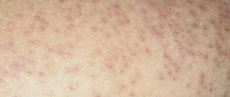
Spider veins (telangiectasias) are small skin formations that look like small dots, lines, and branches, usually red-violet in color. Such formations appear due to local expansion of the walls of the microvasculature structures with the formation of a protrusion. Spider veins on the chest of women not only bring psychological discomfort, but can also be a manifestation of the development of various pathological processes.
Nature of the disease and causes of its appearance
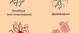
Spider veins on the chest are the result of dilated capillaries that are burgundy or purple in color. The localization of the web on the human body occurs in a chaotic manner, but common places are the face, chest area, arms and legs. The thin capillary web can be of different shapes. In medical practice, the pathology is referred to as rosacea.
Spider veins form due to weakening and expansion of the vascular walls. Poor functioning of the venous valves provokes stagnation of blood flow, which increases pressure on small vessels. Regardless of the place where the “pattern” appears, it is necessary, first of all, to find out the factor that determines its appearance, and then begin to choose a treatment method.
Excessive physical activity in the form of sports can provoke capillary meshwork
It is quite difficult to pinpoint the exact reason for the development of the grid. Its mechanism is a malfunction of the vascular system, accompanied by congestive pathologies. Although in some cases rosacea becomes only a cosmetic defect and affects an absolutely healthy person.
The following can cause the formation of a capillary network:
- hereditary predisposition;
- excessive physical activity in the form of sports;
- vascular diseases;
- hormonal imbalances caused by pregnancy or taking hormonal contraceptives;
- prolonged occupation of a vertical position;
- obesity;
- frequent changes in temperature conditions;
- long exposure to the open sun;
- deep skin peeling, cupping massage.
The formation of a vascular network on the lower extremities can act as a confirmation of varicose veins. The sign becomes especially important for identifying the initial stage of the disease and preventing its development.
If stars appear in the chest area, you should not delay visiting a doctor because of the many reasons for the formation of such a pathology. In addition, capillary networks in the upper part of the body may indicate the onset of an oncological process in the mammary glands.
Spider veins during pregnancy
Pregnancy is a special physiological state of a woman, in which the course of most processes changes. First of all, this concerns hormonal levels, as well as metabolism (metabolism). Spider veins during pregnancy are a consequence of the influence of certain hormones, the level of which increases, on the functional state of the walls of the microcirculatory structures. After childbirth and normalization of hormone levels, telangiectasias do not disappear on their own, but require treatment.
Associated symptoms
The venous network does not pose a threat to human life, but it does not change its appearance in the best way. When pressing on the walls of blood vessels, pain occurs in the damaged areas, redness and itching. The appearance of external symptoms of the mesh is preceded by pain and fatigue in the legs in the late afternoon. Patients often experience leg cramps at night. Further on, a convex mesh of reddish or bluish color is formed, rising above the skin surface. If you press it with the phalanx of your finger, it turns pale, but quickly restores its color.
Diagnosis of the disease
Determining treatment options for a venous network involves conducting an examination to identify the stage, level and volume of vascular pathology. Doppler ultrasound (USDG) is used as the main examination method, but other methods can be used according to indications. In addition to the phlebologist, the examination is carried out by a vascular surgeon, endocrinologist, gynecologist and cardiologist. If necessary, it is possible to prescribe auxiliary examinations to identify pathologies in the functioning of certain organs.
Features of the vascular network on the chest
Most often, stars in the chest area occur in pregnant women. Similar modifications are associated with most conditions, and the formation of the fetus brings changes to the mammary glands. The expectant mother's breasts increase significantly in volume, and the blood supply system at this time begins intensive work in this area. The processes contribute to the appearance of a vascular network.
The mechanism of change occurs against the background of hormonal changes - after conception, a lot of estrogen and progesterone appear in the blood fluid, which directly support the progress of pregnancy. The restructuring of the body is accompanied by painful sensations in the chest area; they disappear after the first trimester of bearing a child.
It is important to choose underwear wisely
You can eliminate the feeling of chest discomfort during pregnancy by following these rules:
- Choosing the right underwear is an important nuance for such a crucial period. A comfortable bra ensures the healthy condition of the mammary glands;
- if pain intensifies or you feel lumps in the chest, you should consult a doctor for proper treatment of the disease;
- During pregnancy, you need to carefully select food products and avoid eating foods that provoke changes in the well-being of the expectant mother.
The use of cosmetic products for stretch marks is permissible only after consultation with the doctor.
The skin surface becomes so dense that barriers to normal blood flow arise. An increase in pressure helps to normalize blood flow, which provokes stretching of the vascular walls and the formation of rosacea. Similar changes are also observed in liver pathologies: hepatitis, cirrhosis, liver cancer. Disruption of the pancreas and thyroid glands can also be accompanied by skin changes that appear on the walls of blood vessels. In such cases, the disease affects not only women, but also men.
What complications are possible after treatment for spider veins?
- Superficial necrosis
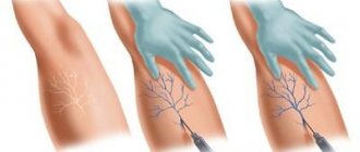
Removal of telangiectasia can be compared to microsurgery. Phlebologists use special magnifying glasses and illuminators for precise injections into the lumen of spider veins on the skin. Such equipment and the experience of a phlebologist make it possible to make an injection precisely into a microscopic vessel, without introducing sclerosant into the thickness of the skin. If the drug is injected intradermally, punctate necrosis sometimes occurs - small ulcers that do not need to be treated and they heal with small whitish scars.
- Deep necrosis
Another dangerous complication of microsclerotherapy can be injection into an arterial vessel. Entry of sclerosant into such a vessel can cause spasm of the cutaneous arteries and major skin necrosis, which may require long-term treatment and may heal with the formation of a rough scar. This is not the phlebologist’s big fault, but the doctor’s experience in choosing a vessel for injection is very important. Upon detailed examination under magnification, it is possible to distinguish a venous vessel from an arterial one and make the right choice. If the puncture site hurts, or if spots or blisters appear in the injection area, you must immediately inform the phlebologist about this condition.
- Microburns during laser treatment and RFO.
These thermal treatments involve heating and welding the vessels. Due to this, a cosmetic effect is achieved. However, in the hands of an inexperienced specialist, laser coagulation can lead to skin burns due to too long exposure to radiation. Most often, the area of this burn is very small and can clinically appear only as a crust, but sometimes there are deeper skin lesions that form rough scars.
- Meshing and hyperpigmentation after microsclerotherapy.
In approximately 10% of patients treated for intradermal vessels, a very thin venous network, called meting, develops in place of the disappeared asterisk. The occurrence of this problem is impossible to predict and very difficult to combat. Metting more often appears after conventional microsclerotherapy without the use of cold; somewhat less frequently, this problem occurs after laser coagulation and RFO. After the appearance of metting, it is better to stop any attempts to combat the asterisks, since practice shows the impossibility of solving this problem with the help of additional sclerotherapy or laser. Over time, this resulting smoky network may decrease on its own or disappear completely.
Hyperpigmentation is not scary, it will definitely go away, but it can take quite a long time. The appearance of the legs after treatment may not satisfy the patient for six months to two years. To prevent this complication, it is necessary to tell the doctor your contraceptive regimen and receive recommendations for contraception during the course of therapy.
In our clinic, a full course of sclerotherapy on the hips begins a month after the first trial session, at which the rate of disappearance of the venous network and the likelihood of complications are assessed. Depending on the results obtained, a decision is made on the timing of the further course of treatment.
- Early relapse of telangiectasis
No patient is immune from the reappearance of new telangiectasis after microsclerotherapy of the lower extremities. The reasons for the appearance of stars on the skin have not been fully established, but the most recognized theory is the postulate of an imbalance of female sex hormones in the body. The levels of these hormones may be normal, but their ratios are different. This is why it is impossible to predict the duration of the aesthetic effect after microsclerotherapy and laser treatment. Relapses are possible, but if these wreaths disappeared well after the first course of treatment, then they will disappear just as well after subsequent ones. In the hands of an experienced phlebologist, complete success in the aesthetic treatment of telangiectasia is usually achieved.
Vascular network on the chest, nuances of occurrence
The venous web does not pose a danger to the body. Such changes can disappear on their own without causing discomfort to the person. However, there are situations of rapid growth of the mesh, which indicates the presence of disorders in the body. Then discovering the true etiology is quite difficult.
Rapid weight loss can cause previously hidden veins to bulge
Additional conditions that cause the development of a venous web in the thoracic region are:
- Powerful physical stress on the body. The appearance of mesh in men can be triggered by taking drugs to stimulate muscle growth.
- Rapid weight loss. A sharp reduction in body weight can cause previously hidden veins to bulge. After gaining normal weight, the formations disappear on their own.
- Surgical intervention. Plastic correction of organs can cause the growth of the venous network.
- Excess weight provokes vascular disorders, as a result of which vascular cobwebs form on the body.
- Pathology of the liver and adrenal glands. The appearance of a mesh under the breast indicates significant disturbances in these organs.
Similar manifestations also occur with high blood pressure - unable to withstand the load, capillaries rupture and hematomas form. If the reason for the changes is known, there is no need to panic. Otherwise, the help of a doctor is required, who will inform you about all the intricacies of the pathology and suggest the correct method of therapy.
Why are varicose veins dangerous?
To this question, most of us will probably answer that spider veins are purely an aesthetic problem. Indeed, you no longer want to wear a short skirt or shorts with veined stars. However, this is not the main thing.
A network of small vessels that appears on the skin may indicate that you are developing varicose veins. In particular, you may already have dilated reticular veins - superficial vessels that have a diameter of about 2 mm and feed the spider vein. In addition, a fine network of red or blue vessels may indicate the presence of chronic venous insufficiency - a set of symptoms that indicates poor circulation in the veins.
Measures to prevent the disease
Venous mesh is formed due to various reasons. If there are no significant disorders in the body, this phenomenon can be eliminated by using cosmetic procedures and taking vitamin complexes. To protect the body from the appearance of unpleasant stars, you must adhere to the following principles:
- complete cessation of bad habits;
- maintaining a balanced diet;
- exercise;
- regular vitamin therapy;
- timely visit to the doctor.
The first thing you need to do is completely give up bad habits.
Taking care of the capillaries is of great importance. Modern drugs allow you to protect them from harmful influences, restore elasticity and firmness. An exceptionally comprehensive approach will ensure care for the body and the disappearance of unpleasant manifestations forever.
At times, the vascular network that forms does not pose a threat to the body. Their appearance may confirm the presence of venous pathologies. In such situations, it is necessary to take care of the condition of the blood vessels, strengthening them. The formation of large sections where veins are clearly visible becomes a significant reason to consult a doctor.
Diagnostics
In order to accurately determine the disease and the degree of its development, you need to make an appointment with a phlebologist. The specialist will conduct an external examination of telangiectasia and palpation of the limbs. As a result, the doctor can draw conclusions about the location of the stars and the nature of the disease.
After this, the specialist will prescribe the necessary studies. They usually include duplex ultrasound scanning, which allows you to assess the condition of blood vessels, determine pathologies and their locations, and assess the direction and speed of blood flow. After this, the phlebologist can draw conclusions about the patient’s condition. For greater accuracy, the doctor may also prescribe tests and studies of other organs, including the heart, thyroid gland, liver, and reproductive system. This will allow us to identify concomitant diseases and establish the cause of the appearance and progression of varicose veins.
Conclusion 3:
You should contact a vascular surgeon immediately after you discover spider veins. Using a series of studies, a specialist will determine the cause of their appearance and prescribe therapy.
Drug treatment
Ascorutin helps strengthen the walls of blood vessels
The early stage of development of rosacea with small foci makes it possible to overcome it with the help of medications. Most often used in therapy:
- venotonics that strengthen vascular walls: Troxevasin, Phlebodia, Venoruton;
- anti-inflammatory drugs that eliminate hyperemia and discomfort: Lyoton, Indomethacin.
Relieves the symptoms of rosacea by strengthening the walls of blood vessels Ascorutin. It is used in the form of tablets or ointment, which is applied topically. The tablets are ground to a powder and mixed with baby cream. The resulting mixture is applied daily to the affected areas.
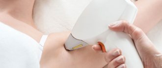
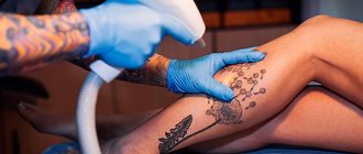
Spider vein removal
The basis of radical therapy for telangiectasias is their removal using various methods of physical or chemical influence, as well as taking measures to prevent re-exposure to the provoking factors of their development. High-quality removal is carried out in our medical center using the following methods:
- Sclerotherapy is the introduction into the lumen of the affected vessel of a special chemical compound (sclerosant), which leads to its gluing.
- Laser therapy is exposure to laser, which leads to “evaporation” of the tissues of the inner surface of blood vessels, followed by gluing.
- Electrocoagulation is the destruction of telangiectasia using local point exposure to electric current of a certain strength and frequency.
- Radio wave surgery – excision of the formation is performed, in which radio wave radiation plays the role of a scalpel.
The choice of method for removing telangiectasia on the chest is carried out individually by a phlebologist-mammologist.