Consultation Services Doctors Results Reviews
Expert tested
Sharubina Karina Petrovna Trichologist, dermatovenerologist, cosmetologist
Publication date: November 16, 2022
Review date: November 02, 2022
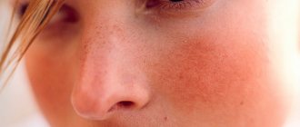
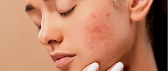
Cuperosis is a condition characterized by impaired blood circulation in the upper layer of the skin. Blood stagnation leads to thinning of the capillary walls due to the heavy load on them. They become fragile and brittle. Capillaries may periodically burst, and the blood coming out of them will be noticeable under the skin. Most often, rosacea occurs on the forehead, wings of the nose, chin, and cheeks. It is usually accompanied by burning or itching of the skin.
In a more advanced stage, rosacea is characterized by the appearance of a vascular network (telangiectasia) that occurs in the area of capillary damage. Couperosis is the initial stage of circulatory disorders, and rosacea is already a chronic skin disease. 10 percent of our planet's population is affected by this disease. At the same time, the disease is much more common in women, although men are more likely to have complicated forms and rapid development of symptoms.
At the first signs of circulatory problems, you should immediately seek help from specialists. After all, treating the initial stages is always easier than the advanced ones.
Avdeyuk Elena Vladimirovna
Dermatovenerologist, cosmetologist
Causes
Since the causes of rosacea can be external and internal, we will consider them separately.
Internal include:
- Rosacea.
- Hormonal surges. Spider veins often appear during menopause or pregnancy.
- Diseases associated with the thyroid gland, pancreas, ovaries.
- Some types of cardiovascular diseases, as well as gastrointestinal tract diseases.
- Selected autoimmune diseases.
As already mentioned, all of these factors are quite rare. Couperosis often appears for external reasons, which include:
- Regular exposure to UV rays.
- Bad habits. Alcohol and smoking have a strong effect on blood vessels.
- Improper skin care. If the blood vessels are weak and the skin is damaged by scrubs or aggressive acids, the symptoms of rosacea will intensify over time.
- An unbalanced diet, which contains a lot of spicy, too hot food, as well as chocolate products and coffee.
- Severe temperature changes when the skin is first exposed to heat and then to cold.
Often it is not possible to determine the specific cause of rosacea, because its first symptoms begin to appear at a young age - the patient simply does not remember what could have influenced it.
Etiology and pathogenesis of rosacea
The etiological factors of rosacea can be divided into endogenous and exogenous.
Endogenous factors include the influence of heredity. A decrease in the elasticity of the vascular wall is a genetically determined condition, which can be aggravated by various provoking (exogenous) factors - heat, cold, hormonal imbalance, somatic diseases, weakness of the valve apparatus of the veins of the lower extremities, etc.
Among the exogenous factors, it is worth highlighting the following:
- Nervous strain - in this case, large main vessels narrow, which increases the pressure in them, and the vessels of the peripheral network, on the contrary, expand.
- Smoking - promotes spasm and decreases the strength of the vascular wall.
- Diet - too hot, spicy or salty food, frequent and/or heavy consumption of alcoholic beverages provoke exacerbation and progression of rosacea.
- Climate - this includes ultraviolet radiation, prolonged exposure to strong gusty winds, cold or too hot and humid climate.
- Lifestyle - rosacea can worsen after visiting a sauna or bathhouse, with mechanical pressure on the skin (wearing tight synthetic clothing), while working with formaldehyde, etc.
Food triggers for rosacea are similar to those for rosacea. This includes hot tea and coffee, wine, strong alcohol, products with capsaicin (spices, hot sauces, hot and red peppers) and with cinnamaldehyde (tomatoes, chocolate, citrus fruits).
There is evidence that disruption of the microbial composition of the skin can provoke dysfunction of the epidermal barrier, which increases the skin's sensitivity to external factors and contributes to the development of rosacea.
Symptoms
Although this disease can affect any part of the skin, it most often manifests itself on the face - on the wings of the nose, cheeks and chin. This is due to the fact that in these areas the skin is thin and elastic.
This type of vascular network does not hurt, and there is rarely a feeling of itching or burning. It happens that after the appearance of such stars, the process slows down and stops - if you do not injure the skin and care for it properly, this does not cause any discomfort to the patient (or only aesthetic discomfort). But in some cases, the disease progresses and becomes more serious: the affected areas of the skin begin to turn blue, the skin or limbs (if rosacea is on the legs) swell, and inflammation begins.
Even if physically the symptoms of rosacea do not bother you, but it has appeared recently, you need to consult a dermatologist: if possible, find out the reasons for the deviation and understand whether there is any additional danger in it.
Mesobotox against rosacea
Botulinum toxin, getting under the skin, begins to affect the hormone serotonin, as well as ion channels responsible for the appearance of many signs of rosacea (the effect of dilated blood vessels) and its chronic form - rosacea.
This allows you to normalize neurovascular disorders and, thereby, reduce the severity of redness and uneven skin texture. Botulinum toxin also has the ability to reduce the hyperactivity of subcutaneous vessels and normalize blood microcirculation in them. This leads to a decrease in persistent erythema and a significant decrease in the intensity of redness as a result of sudden flushes of blood to the face, which often occur in patients with rosacea during physical activity, stress, exposure to hot water, wind, frost and other negative factors.
Stages on the face, complications
It is customary to distinguish four stages of rosacea:
- 1st. As a rule, the vessels do not appear yet or there are only two or three of them - a person simply often experiences redness in the face or other parts of the body. After severe redness, the skin can sometimes dry out (more rarely, peel off).
- 2nd. The redness of the cheeks is more obvious (often when washing or touching), the vascular pattern is more active. Some patients have a tingling or burning sensation.
- 3rd. The redness in the affected areas does not disappear anywhere - it is constantly on the skin. Severe dryness of the skin is observed, and an inflammatory reaction may begin.
- 4th. All facial vessels are already involved in the process; there is a spasmodic effect, due to which certain areas of the skin become pale.
The health and life of the patient with this disease is not in danger. But it is important to understand that rosacea is the enemy of young skin. The aging process begins to develop more actively, the skin loses its attractive appearance. This has different levels of significance for patients of different ages. If the problem occurs in adulthood and old age, many simply adhere to the minimum recommendations of doctors and do not pay too much attention to it.
But rosacea in children and young people requires a more serious approach - such patients still have a whole life ahead, so the aesthetic aspect is also very important for them.
In addition, it is important to remember that in some cases this disease still indicates problems in the body that can be dangerous. And in order to avoid problems, it is necessary to get advice from specialists.
Diet for rosacea
Can
- Fermented milk products (cottage cheese, kefir, milk, low-fat sour cream, fermented baked milk);
- Lean meats (chicken, turkey, veal), preferably steamed or roasted
- Vegetables, especially all types of cabbage, zucchini, squash - preferably eaten raw or semi-raw
- Fruits (hypoallergenic)
- Berries (hypoallergenic, not too sour)
- Long-term carbohydrates (cereals: buckwheat, oats, barley).
- Vegetable oil (any in small quantities)
- Dried fruits (as dessert)
- Greenery
- Fish (low-fat types)
- Rye or whole grain bread
Must be excluded
- Fatty meats
- Spices
- Snacks (chips, crackers)
- Smoked products
- Pickles
- Light carbohydrates (cakes, pastries, buns, any other types of baked goods, sweets)
- Coffee and strong tea
- Canned foods
- Alcohol
- Fast food
Diagnostics
To determine rosacea on the body, a dermatologist does not need much research. But it is important for patients to understand that they cannot do this on their own; it is necessary to differentiate this condition from others that may be more dangerous.
For diagnostics the following are used:
- Examination, history taking and dermatoscopy.
- General urine and blood tests, biochemical blood tests, as well as tests for certain types of hormones.
- Ultrasound of the thyroid gland and abdominal organs (optional).
If a specialist suspects hidden diseases, this list may expand.
Perioral dermatitis
Synonyms: rosacea-like dermatitis, flight attendant disease - rashes around the mouth: redness, swelling and papules.
Diagnostically difficult to distinguish disease of unknown etiology. Provoking factors : the use of steroids both locally and inhaled, fluoride-containing pastes, occlusive cosmetics.
The main diagnostic differences from rosacea: bright erythema and telangiectasia are rare. Localization: nasolabial folds, perioral region, chin, lower eyelids. Women aged 20-30 years and children are affected.
Distinctive feature : rapid regression of rashes, if a trigger factor is identified.
If visual diagnosis is difficult, a histological examination is recommended.
Treatment
If there is an underlying disease that has caused rosacea on the face or body, then treatment begins with its elimination. In this case, the patient is often accompanied not only by dermatologists, but also by endocrinologists, cardiologists, and gastroenterologists, depending on the disease underlying the problem.
When the disease is independent, in the first and second stages, doctors usually recommend proper skin care and monitoring your condition. The third or fourth stage already shows the treatment of rosacea by removing dilated vessels using one of several methods:
- Ozone therapy. It involves the introduction of a certain mixture into the lumen of a damaged vessel.
- Laser therapy. A laser beam is used to narrow blood vessels (the best option is a neodymium laser).
- Phototherapy. When using it, blood vessels are restored using photo flashes.
- Electrocoagulation is essentially cauterization of blood vessels with electric current.
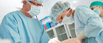
If there are no additional diseases, this problem can be solved quite effectively on an aesthetic level. For detailed advice, we recommend contacting the dermatologists of Meditsina JSC in Moscow - the clinic’s doctors will help patients at any stage of the disease.
Seborrheic dermatitis
Seborrheic dermatitis is a chronic inflammatory disease associated with a violation of the amount and composition of sebum and a disturbance of the skin microbiota.
Clinical manifestations : hyperkeratosis, peeling, seborrhea, inflammation and itching.
Localization : areas with the largest number of sebaceous glands, including the T-zone of the face, as well as the scalp, upper third of the back and chest area, ears.
The common feature is centrofascial erythema, the distinctive features are: with rosacea – papules and telangiectasia, with seborrheic dermatitis – the formation of scales and peeling, which are not characteristic of rosacea.
Prevention
To avoid having to carry out complex treatment of rosacea on the face, you must adhere to the following measures:
- Give up bad habits, switch to proper nutrition.
- The right way of life. Healthy sleep, moderate physical activity, avoiding stress - all this is very important.
- Proper skin care. It is best to consult a cosmetologist who will select care taking into account your characteristics.
- Regular skin protection from the sun. There is no need to be outside during periods of particularly sunny activity (from twelve in the afternoon to five in the evening), and it is also important to always use sunscreen.
- Protecting the skin from hypothermia - there are also special nourishing creams for winter.
Since the problem is related to the quality of the walls of blood vessels, a very important factor is proper nutrition. Once every year or two, for this reason, you need to visit a nutritionist-endocrinologist, take tests for missing substances and take a course of vitamins, and also adjust the menu in accordance with the doctor’s recommendations.
The whole truth about spider veins
Spider veins are not a disease!
So, spider veins or, as they are often called capillaries (telangiectasis), are not exactly a disease, but rather a cosmetic problem! This is the honest truth, which is often passed off as varicose veins.
Yes, on the one hand it is varicose veins, but varicose veins are at the level of skin vessels - capillaries and intradermal veins. Such microvaricose veins do not cause any painful symptoms, pain, cramps, swelling, or blood clots. Nothing. Only aesthetic discomfort.
Patients who often come in with pain in the legs, swelling, and discomfort in the calves notice spider veins on their legs and place all the blame on them. And therefore, instead of “treating” with a newfangled “anti-varicose” gel in the area of blood vessels, we recommend looking for the real cause of your discomfort in the condition of your legs (meaning symptoms) - for example, “heavy leg” syndrome, flat feet, chronic venous insufficiency, spinal osteochondrosis, etc. d.
Spider veins are not a disease, which means there is no need to strengthen the blood vessels!
Strengthening blood vessels with medications and gels is a waste of time. You can train muscles, ligaments, and develop endurance. But the blood vessels cannot be strengthened even with the help of medications.
What do we know about spider veins (webs)?
The problem is cosmetic and widespread, spider veins appear at different ages in approximately 70% of women due to the fact that female sex hormones are synthesized in the female body, starting from adolescence, and this stimulates the translucency of intradermal veins, and with age, the appearance of such unwanted elements like spider veins.
For example, a 9-month pregnancy and the accompanying changes in a woman’s body at the hormonal level, as a rule, aggravate the problem. This is normal and physiological, and most importantly, harmless.
That is, you can be calm about your health.
Is there a way to prevent the appearance of unwanted vascular elements?
You can read for hours on the Internet about preventing the appearance of ill-fated blood vessels. And even watch it on TV in popular talk shows. This is a good business - the problem is very common, and if you come up with some kind of “miracle remedy”, many will definitely try it and buy it. There used to be a drug called Asklezan - both tablets and gel. It was necessary to smear and drink 4-6 courses and they supposedly became smaller or “faded” or decreased. In general, they wrote about it in newspaper advertising, people ran and bought it. They smeared themselves. Well, of course, the effect is zero.
There is no way to prevent the appearance of spider veins!
They will gradually appear at different rates in different periods of life.
And if you don’t like it, you can think about treating existing unaesthetic telangiectasis, but it is impossible to slow down or stop this process, even if you take courses of venotonics and daily smear the area of the spider veins with various venotonic creams with or without leeches. This will not affect the process of their appearance and progression in any way.
Treatment of spider veins with medications is useless!
The basic concept of treatment for telangiectasis is not treatment of the whole body or strengthening of blood vessels or drug support by introducing any drugs into the area of spider veins using gels - this is a waste of time. Not a single star will fade, not a single one will disappear. It is a fact. All we can do is inject a sclerosant into the vascular element so that it damages the inner layer of the asterisk and it closes (sclerotherapy, or rather microsclerotherapy) or electrocoagulate the vessels every millimeter or percutaneously treat with a laser again every mm.
That is, the main concept of treatment is to destroy the vascular element in one way or another without damaging anything around it.
Percutaneous laser coagulation
Special lasers are used to treat telangiectasis. The basic principle of treatment is this: a laser beam passes through the skin and is absorbed by the red blood in the vessel, after which the blood heats up and explodes. The vessel is “brewed”. This is a very good method, but only for small and very superficially located meshes, for example, on the face and torso. On his feet he works very unsteadily and it is difficult to get results. Used in Russia mainly by cosmetologists.
In my opinion, a significant disadvantage of percutaneous laser coagulation for leg telangiectasis is the limitation in the diameter of the vessels, the high pain of the procedure and the high probability of difficult to predict side effects (burns of the skin surface, depigmentation in the affected area).
Electrocoagulation of capillaries
Using a very thin needle-electrode, many injections are made into the capillaries with simultaneous exposure to electric current. The procedure is painful and requires anesthesia with a skin anesthetic cream (EMLA on the legs takes effect after 2 hours of exposure under cling film). Therefore, you either smear yourself with Emla cream at home and wrap your legs in film and come to the doctor, or you receive hundreds of rather painful injections. The frequency of injections into the capillary is every 1-1.5 mm. That is, 1 cm of capillary will require 6-10 injections.
Sclerotherapy is the method of choice
Sclerotherapy is an injection technique for the treatment of spider veins and reticular veins. It is most effective against small veins and capillaries. Its meaning is to introduce a sclerosant into the lumen of the vessel, which causes chemical damage to the inner layer of the vessel, followed by its “closure”. After the vessel has closed, it begins to gradually dissolve (several weeks - several months).
Today, sclerotherapy of reticular (intradermal veins) and capillaries is called microsclerotherapy and has many modifications; moreover, everyone performs it differently, so the results can also be very different.
Types of microsclerotherapy
There is a technique of liquid sclerotherapy, when the sclerosant is administered in the form of a solution and this works well on small capillaries with low-intensity blood flow, and a technique of introducing foamed sclerosant (foam-form sclerotherapy), when air or other gas (CO2, for example) is added to the sclerosant, whipped by distillation through an adapter from one syringe to another and after 10-20 strokes a stable foam is obtained.
The sclerosant foam introduced into the lumen of the vessel expels blood well and is well retained there. Foam-form sclerotherapy works better on vessels with more intense blood flow.
Liquid sclerosant works more intelligently and gently, with a minimum of side effects, foam is more aggressive. In the same treatment area, liquid sclerotherapy can be used for some veins and capillaries, and foam for others. A phlebologist can use various scleropreparations, of different concentrations, in liquid or foam form during one session on one treatment area. It all depends on his experience, working style, etc. This is about the active advertising of the miracle method of foam-form sclerotherapy, which is separately positioned as a newfangled and very effective method. We've been using foam for about 10 years and it works great in certain situations, but not much else, especially with mesh.
Microsclerotherapy - a special approach
When it comes to sclerotherapy for spider veins, it is necessary to immediately clarify the situation.
This is an intelligent problem and requires a certain doctor’s experience and a certain technological approach.
It is necessary to take into account the volumes of the administered drug and its concentration. On the other hand, today microsclerotherapy is a methodologically verified procedure, and if everything is done according to technology, then the results will be as expected.
What patients don't understand
First of all, after sclerotherapy sessions, aesthetic results will not be immediate, but after some time. The fact is that closed vessels are visible through the skin and are noticeable. That is, if after the injections the spider vein does not close, then it does not change its color and remains the same as it was; if it closes, it darkens (the remaining small amount of coagulated blood shines through the skin).
The entire time that sclerotic telangiectasis is resolving, it will be noticeable. As soon as it dissolves, the marks will disappear.
This applies not only to sclerotherapy, but to any other effective treatment method. And it is important to understand this before you decide to undergo sclerotherapy. It is necessary to choose the right time for treatment so that you have a reserve of time for aesthetic rehabilitation.
How to get rid of spider veins?
It is clear that you are reading this to understand how you can improve the aesthetics of your legs. Considering the fact that in most cases the method of choice for primary treatment is sclerotherapy, it makes sense to see a phlebologist. An ultrasound of the venous system will be performed and treatment will be recommended. Naturally, in cases of small red capillaries, percutaneous laser coagulation (PLC) may be recommended to you, but this is unlikely, since PLC is usually used for follow-up treatment (only small red capillaries) and usually by cosmetologists.
In most cases of reticular varicose veins, the treatment area includes reticular (feeding) veins (blue), large and medium capillaries (purple) and small ones (red).
In order for the treatment to be effective, the phlebologist begins with the reticular (feeding) veins (marked with blue arrows in the photo), then moves on to large capillaries, then to medium and small ones. Of course, only red vessels can be treated, but practice shows that this is ineffective if the feeding reticular vein remains.
Stage one - sclerosing the “feeding” reticular veins
Often reticular veins communicating with telangiectasis are clearly visible without any equipment (magnifying glasses, polarized light). However, at the same time, poor results of sclerotherapy are associated with poor visualization of these very feeding veins, which can be obscured by the spider veins located above them. A solution to this problem has been proposed by companies that produce Veinviewers - devices based on the thermal imaging effect, which allow brilliant visualization of intradermal veins.
The phlebologist begins the session under the control of Veinviewer, punctures the feeding vein and closes it by injecting a sclerosant.
This is a simple and effective technology that we have been using for over a year to more successfully treat spider veins. Moreover, Venovisor makes it possible to clearly see the vein from which the unwanted vascular element is filled. Veinviewer technology is the key to understanding the problem of spider veins.
Stage two - sclerosing spider veins (telangiectasis)
This is a very simple manipulation at first glance - injecting the drug into the spider veins. The vascular network is punctured with a very thin needle and a small amount of sclerotherapy is injected into it. The introduction is slow and very small in volume.
This allows you to avoid long-term side reactions (matting (see below), pigmentation). Sclerotherapy can be most successful with multiple injections and small amounts of sclerotherapy.
After the introduction of sclerosant, we observe the redness of the element and its reaction. There may be a slight tingling sensation in the injection area. Thus, in the first session, as a rule, the feeding reticular veins are treated, in the second and subsequent sessions - telangiectasis (spider veins).
A few words about haste in treatment
You come 2-3 months before your vacation with the desire to get rid of capillaries, but you did not know that you cannot rush in the treatment of spider veins.
A number of side effects, which can then subside for months and years, are associated not only with the sclerotherapy technique, but also with short breaks between sessions.
Ideally, a session on one leg in the same zone should be carried out at intervals of 3-4 weeks. This is the optimal schedule. That is why we recommend treating spider veins as planned, taking into account your vacations and beach holidays and “much in advance.”
How to make injections painless and improve the results of sclerotherapy?
In microsclerotherapy, injections are performed with cosmetic needles with a diameter of 0.3 mm. These are very thin needles and the injection is insensitive. However, some patients are afraid of injections and for them this type of treatment creates psychological stress. We have solved this problem because we perform cryosclerotherapy. The technique was born in the USA and, with the light hand of the well-known Luis Navarro, was called painless sclerotherapy.
This new method involves cooling the skin for a few seconds with a blast of cold air before injection, creating cold anesthesia of the skin. This is enough so that the patient does not feel the injection.
The miracle of cryosclerotherapy
As it turned out later, this is not the only positive thing from cryosclerotherapy - bruises, hematomas, pigmentation and other side effects after the session are much less than after conventional sclerotherapy. This is due to local vasospasm in the treatment area. And, as a result, in spasmodic vessels the sclerosant works more effectively with a lower concentration and a smaller injected volume.
Compression and sclerotherapy
It has always been believed that round-the-clock compression is necessary for the entire period of sclerotherapy - during the day with stockings, at night with an elastic bandage, plus another 2 months after the end of treatment. That is, only six months. For many patients this was unbearable. But if this is true, then only for sclerosis of large veins. When performing microsclerotherapy of spider veins and reticular veins, compression is usually needed until the evening - either a bandage or stocking. The main purpose of compression in such cases is to reduce the formation of bruises after the session. In rare cases, the daily compression regimen can be extended to 3-5 days.
Modern technology for microsclerotherapy allows this type of treatment to be carried out with virtually no compression and at any time of the year, even in summer.
Innovations in sclerotherapy
Globally, the microsclerotherapy technique has not changed. Same drugs, same injections. Foam-form sclerotherapy, when used on spider veins, usually does not live up to expectations, so sclerotherapy with a liquid solution is preferable. However, since 2013 we have introduced a couple of innovations in technology: cooling the skin during the procedure and the use of a Veinviewer.
These improvements make the procedure more comfortable, reduce the number of side effects, and improve control over the administration of the drug.
Possible side effects of microsclerotherapy
Pigmentation
The most common side effect of microsclerotherapy is pigmentation at the site of sclerotic veins and spider veins. After administration of the sclerosant, the inner layer of the vessel is damaged; at first the vessel disappears, but after a few minutes it fills with blood again. If the vessel subsequently thromboses (closes), then a black tar-like mass (coagulum) is formed in it, which is darker than blood and the vessel shines through the skin brighter than the original one. Over time, the closed vessel with coagulum dissolves. Usually this is several months. If this process is long and the coagula is quite pronounced, then iron is deposited in the skin and it becomes light brown. Actually this is called pigmentation. In most cases, pigmentation goes away within six months, but in 5% of cases it can take a year and in 1% of cases up to 2 years. But it always passes, which makes me happy.
The severity and duration of pigmentation after sclerotherapy is individual and depends on the characteristics of pigment metabolism in the patient’s body.
Matting (neovascularization)
Matting is a reaction in the form of the appearance of small capillaries in the treatment area. This is a complex and sometimes difficult to explain and predictable phenomenon. It is largely determined by hormonal levels (manifested in women), as well as by the individual reaction to the administration of sclerosant. Most often associated with the use of high concentrations of scleropreparation or a large volume of administration.
That is why you should not try to inject all the vessels at once in the first session - the dose may be large and this will cause an undesirable reaction.
Most often, matting occurs if sessions on one leg occur very often (once a week, for example). A more gradual style of treatment with longer breaks produces better results. You should also not conduct more than 3 sessions in a row on one leg without a long break (3-6 months).
How to minimize the risk of side effects during sclerotherapy?
Introduce a small amount of sclerosant through multiple injections, take long breaks, and take a long rest after 2-4 sessions. Sclerotherapy should be like the leisurely work of an artist who gradually draws the details of a picture.
That is why it is necessary to approach your treatment without haste and without any limiting circumstances. Choose a time and go to the procedures calmly.
#FORM_PSK1#
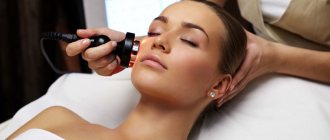
Laser coagulation method
The essence of the procedure is that the laser seals the walls of damaged vessels, which prevents the flow of blood into them, due to which the network of capillaries becomes completely invisible.
The advantages of the technique are its painlessness, the absence of any marks on the skin after the course of treatment. In addition, I note that this does not cause any harm to the entire circulatory system.
Sessions can be held both in winter and summer. The duration of one procedure is about forty minutes. The number of appointments with a cosmetologist in this case will depend on the degree of complexity of the disease. At the initial stage of rosacea, four sessions may be enough, but to get rid of advanced rosacea, it is best to spend at least seven.
It is important not to use any cosmetics during the treatment period; the effect of such therapy will be noticeable on the third or fourth day.