| Appointment with a dermatologist at the clinic. Call a dermatologist at home. | Reception is strictly by appointment, make an appointment by phone: +7 | Prices for services | Reviews about the clinic |
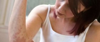
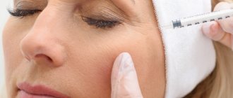
The skin is the largest human organ and the most sensitive to changes in the body. If there are malfunctions in the functioning of internal organs or if there is an adverse effect on the body, the skin reacts with rashes. This symptom must be taken seriously. You should visit your doctor without delay.
It is important to find out whether the rash is infectious or allergic in nature. Both require timely treatment, and sometimes isolation of the patient if the disease is contagious. Our clinic offers the services of experienced specialists, as well as sufficient facilities for modern, accurate laboratory and diagnostic tests.
Make an appointment with a dermatologist by phone or by filling out the online form
| Select a clinic | Demodicosis | Rash on shoulders | Calling a dermatologist to your home |
Answers to frequently asked questions about skin rashes:
- Which doctor should you contact for a skin rash?
- Is the skin rash contagious?
- What diet is necessary for skin rashes?
- What diagnosis is needed for a skin rash?
- Why is a skin rash dangerous?
- Why is it necessary to get tested for a skin rash?
- What diseases does a skin rash indicate?
- What examination is necessary for a skin rash?
- Which skin rash is dangerous?
- How to distinguish an allergic rash from an infectious one
- How to get rid of skin rashes?
- How to get rid of itching skin rash?
- What organs are affected by a skin rash?
- How to prepare for an appointment with a dermatologist?
- How to get checked for skin diseases?
- What diseases does a dermatologist treat?
- What tests should be taken by a dermatologist?
- What diagnostics can a dermatologist perform in the clinic?
- Where to go with a skin disease?
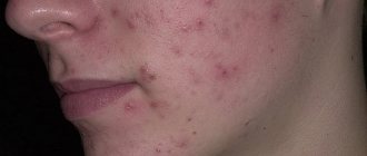
Symptoms of acne in papulopustular form
Acne vulgaris in the papulopustular form manifests itself in the form of papules and pustules. These acne elements have their own differences18,97:
- Papules. Small inflammatory nodules of a conical or hemispherical shape. Their diameter is 2-4 mm. They have a bluish-red color and elastic consistency. Their full development cycle is about 10-12 days. After their resolution, post-inflammatory spots may remain.
- Pustules. Inflammatory nodules, inside of which a cavity is formed filled with pus. They, like papules, have a red-blue color. Over time, the purulent contents dry out and a crust forms on the surface. After its removal (intentional or spontaneous), a trace remains on the skin - post-inflammatory hyperpigmentation.
Pustules can form on their own or transform from papules.
What is a pustule?
A pustule is the primary inflammatory element of a rash, which is a purulent process in the dermis or epidermis. Inside the pustule is pus, which may be gray or white. If the pus has a greenish or yellow tint, then the pustule is a consequence of infection. Sizes range from one to 10 millimeters. Typically, painful redness can be observed around the purulent head. By nature there are follicular and non-follicular. Follicular pustules form in the hair follicle, hence their name. Non-follicular - develops outside the hair follicle. Pustules also differ in shape: cone-shaped, flat, spherical. Sometimes when you press on a pustule, it may be accompanied by a painful reaction.
Why do papulopustular acne appear?
Heredity influences how acne develops and progresses. There is a genetic predisposition to hyperandrogenism, hypersensitivity of the sebaceous gland receptors to androgens, increased activity of 5-alpha reductase, which is involved in the conversion of testosterone to DHT. It is DHT that helps increase sebum production.29
Inflammation in acne can be either aseptic, developing in sterile conditions, or reactions to substances produced by propionibacteria. 29
Clindovit® gel is a topical antibiotic. Its main active ingredient is clindamycin phosphate. The drug exhibits antimicrobial activity against propionibacteria.6
The causes of acne can be associated with diseases of internal organs and systems, for example, the gastrointestinal tract; rashes can be a consequence of dysbacteriosis, hypovitaminosis, metabolic disorders, and decreased immunity. 29
The causes and treatment of acne are interrelated. So, if the rashes in women are of a hormonal nature, in addition to topical therapy, oral contraceptives may be prescribed. 29
Diagnostic methods
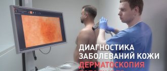
If papules, pustules, or vesicles appear, it is recommended to visit a dermatologist, consult with an infectious disease specialist, venereologist or allergist. At the appointment, the doctor interviews the patient, examines the skin using a dermatoscope, and prescribes a series of examinations.
The following diagnostic techniques are used:
- liver tests;
- stool analysis for worm eggs;
- skin allergy tests;
- bacterial sowing;
- biopsy;
- general and biochemical blood tests;
- chest x-ray;
- HIV test;
- plasma analysis for hormone levels;
- skin scraping microscopy;
- urine test.
Treatment of papulopustular acne
Treatment of papulopustular acne depends on the severity of the disease. If there are many comedones and only a few papules on the skin, most likely the patient has mild acne, for which topical medications are prescribed. If there are more than 10 papules and pustules, this indicates an average severity, for which treatment should be combined, including systemic medications. 18
Clindovit® gel is intended for the treatment of mild to moderate acne. It should be applied to previously cleansed skin 2-3 times a day.6
In addition to medication, there are other methods of treating acne. Thus, the use of laser and phototherapy is considered effective. Ozone therapy is also used. But each of these methods has its own contraindications, which in most cases include the inflammatory process. With the help of drug treatment, it is possible to achieve remission, and hardware methods will help prolong it and improve the condition of the skin.
Diseases that occur with pustular rashes on the palms and soles include palmoplantar psoriasis, palmoplantar pustulosis, acrodermatitis persistent pustular Allopo, and Andrews pustular bacteridus. There are disagreements among researchers in the interpretation of the concepts palmoplantar psoriasis and pustulosis. In some sources, palmoplantar pustulosis is classified as a separate nosological form, or is considered a type of palmoplantar pustular psoriasis [1]. There are localized pustular psoriasis of the Barber type (syn.: pustulosis of the palms and soles; chronic palmoplantar pustular psoriasis; persistent palmoplantar pustulosis; pustular psoriasis of the extremities). Many domestic scientists are of the opinion that various forms of pustular psoriasis are nosologically independent [2, 3]. At the same time, the question of whether non-infectious pustular dermatoses belong to pustular psoriasis continues to be discussed. Some domestic scientists [4] believe that in terms of clinical and morphological manifestations, palmoplantar pustulosis is closer to Andrews bacterid, and not to psoriasis. Some foreign authors [5] also consider palmoplantar pustulosis as a separate nosological entity. Data from genetic, immunological, and histological studies allow dermatologists to prove these statements. Perhaps this is due to the different meanings attached to the concept of “palmoplantar pustulosis”. In the generally accepted ICD-10, the form palmoplantar pustulosis (L40.3) is distinguished, but it is considered under the heading of psoriasis (L40), as well as acrodermatitis persistent Allopeau (L 40.2). With acrodermatitis persistent Allopeau, pustular rashes of a different localization may appear, sometimes similar to generalized pustular psoriasis, which also indicates that this disease belongs to psoriasis [5]. This issue is also important in terms of differential diagnosis, since errors in diagnosis and patient management tactics often occur. Damage to the palms and soles causes a lot of physical and emotional suffering for patients, disrupting performance, interpersonal relationships, and affecting the quality of life.
Localized pustular psoriasis (Barber type), a type of pustular psoriasis characterized by erythematous-squamous and pustular elements in the palms and soles [1], is widespread throughout the world, more common than the generalized variety of pustular psoriasis [6, 7], but significantly less common than psoriasis vulgaris. The disease occurs in the period from 20 to 60 years, mainly at the age of 40-50 years, very rarely after 60 years and only in 19% of cases before the age of 20; in women 3 times more often than in men [1], while psoriasis vulgaris more often affects men [5]. A family history of this form of psoriasis is noted only in 7-24% of cases; the connection with antigens of the HLA system has not been established [1]. Analysis of some genetic studies [5] allows us to assert the nosological independence of palmoplantar pustulosis and psoriasis, but other studies [8] do not allow us to identify clear differences between these pathologies, which coincide in many aspects. According to the Tyumen Regional Dermatovenerologic Dispensary for the period 2008-2012, limited palmoplantar psoriasis accounts for only 1.4% of the total number of registered cases of vulgar psoriasis and 2% of newly diagnosed cases of psoriasis (Fig. 1, 2)
.
Figure 2. Patient K., 51 years old, with a smoking history of 35 years.
Clinical manifestations of palmoplantar psoriasis. Symmetrical lesions of the skin of the hands (a) and feet (b). The exact cause of localized pustular psoriasis is unknown. Provoking factors are foci of focal infection, primarily odontogenic and tonsillogenic [1], Helicobacter pylori
, as well as stress, anxiety, the use of certain medications, and liver dysfunction [2]. Sometimes the disease first appeared after long-term treatment with lithium drugs. Smoking and unfavorable environmental factors (high humidity and temperature) aggravate its course, and a more pronounced association has been identified between palmoplantar pustular psoriasis and smoking, especially in women [1, 8]. Smoking cessation was an important treatment intervention given the observed abnormal response to nicotine [5].
Localized pustular psoriasis occurs with or without psoriasis vulgaris rashes, most often in patients with a personal or family history of psoriasis, although often independently [1].
A possible mechanism for the formation of pustules in pustular psoriasis is considered to be a decrease in the activity of antileukoprotease in the skin as a result of a protease/antiprotease imbalance [5]. To date, extensive material has been accumulated on the significant role of infectious agents in the occurrence, spread and activation of the psoriatic process [6]. The contents of the pustules are sterile, although some authors, in particular G.Ya. Sharapova et al. (1983) noted the presence of Staphylococcus aureus in some patients. According to numerous studies [7], microorganisms that sensitize the patient’s body against psoriasis are often streptococci and Staphylococcus aureus, as well as foci of chronic infection. Staphylococcus aureus and streptococci secrete exotoxins that play the role of superagents capable of binding to proteins of the major histocompatibility complex of tissues on antigen-presenting cells, keratinocytes, T-lymphocytes, monocytes, which leads to the formation of cytokines that enhance the proliferation of keratinocytes [5].
Subjective symptoms boil down to burning, itching and a feeling of discomfort in the palms and soles; often these sensations precede the appearance of fresh elements. Cases when in the early stages the disease is represented by single plaques or is asymmetrical in nature are rare [1]. With severe severity of rashes, the quality of life of patients is significantly reduced due to pain, inability to stand, walk, or work with their hands [5].
Clinically, on a scaly erythematous background with sharp boundaries, there are pustules, which, unlike generalized pustular psoriasis, are located deep in the epidermis, which is due to the morphological structure of the skin of the palms and soles. Localized pustular psoriasis is manifested by sterile yellow pustules with a diameter of 2-5 mm, approximately the same size, clusters of which within a few hours appear symmetrically on the apparently healthy skin of the palms, mainly in the thenar area, less often - the hypothenar, even less often - the middle and distal part of the palm and soles, mainly in the area of the arch of the foot [1]. Subsequently, a halo of hyperemia appears around individual elements. New rashes are associated with places of favorite localization and rarely spread to the back of the hand and foot, wrists, and fingers. The lesion is usually symmetrical, but a unilateral arrangement of elements may be observed. Sometimes the pustules spread to the dorsal surface of the fingers, toes, or the inside of the wrist. The skin of the terminal phalanges of the fingers is usually not affected. The disease lasts for years, with off-season exacerbations followed by remissions [9]. Episodes of new pustular eruptions occur over varying periods of time and are strictly limited to their preferred locations. Occasionally, lesions of ordinary psoriasis appear, but more often the rash remains localized [10].
Subsequently, the color of the pustules changes to yellow-brown and dark brown. Without opening, the pustules dry out to form brown crusts, which resolve within 8-10 days. The presence of pustules at different stages of evolution (evolutionary polymorphism) gives the lesion a multi-colored color. Later, the pathological process manifests itself as sharply defined erythematous-squamous plaques, within which and along the periphery there are multiple yellow and brown pustules, some of which merge to form “purulent lakes” [1]. Remission begins with a decrease in the number of new pustules, although the skin of the palms and soles may remain erythematous, erythematous-squamous, or hyperkeratotic, clinically resembling eczema. Remissions continue for weeks or even months [11]. Thus, unlike other forms of psoriasis, the course of pustular psoriasis of the palms and soles is chronic. According to W. Enfors and L. Molin [1], at the time of examination 10 years after the diagnosis of pustular psoriasis of the palms and soles, its manifestations were absent in only 28% of patients.
Localized pustular psoriasis is often associated with hyper- and hypothyroidism, diabetes mellitus, arthropathy; it is often diagnosed in patients with SAPHO syndrome (synovitis, acne, pustulosis, hyperostosis, osteitis), namely with chronic recurrent multifocal osteomyelitis, lesions included in this syndrome sternoclavicular and sternocostal joints, pustular arthroosteitis, peripheral arthritis, pseudo-infectious arthritis involving the sacroiliac joints in the pathological process [1, 5].
In the differential diagnosis, fungal, scabies, eczematous dermatitis, and Andrews pustular bacterium are excluded.
The diagnosis of pustular psoriasis of the palms and soles is established on the basis of the characteristic clinical picture and course of the disease, and in difficult cases should be confirmed histologically.
Andrew's pustular bacterium
(syn.: Lever's pustulosis of the palms and soles) is a type of pustulosis of the palms and soles, which some authors classify as localized pustular psoriasis [1, 5]. A.A. Kalamkarian et al. [12] deny the existence of this disease as a nosological form. This variant of pustulosis was isolated by G. Andrews in 1934 and is characterized by the formation of small pustules with sterile contents on the unchanged skin of the palms and soles.
The etiology and pathogenesis are not clear, but since the occurrence of the disease is facilitated by foci of chronic infection in the body, especially chronic tonsillitis [1], chronic recurrent osteomyelitis, osteoarthritis [7], an infectious-allergic origin of the process is assumed, or more precisely, a hypersensitivity reaction to streptococcal antigens.
Clinically, the disease manifests itself as multiple blisters and pustules on the palms and soles. The rashes, as a rule, are symmetrical, starting from the center, gradually covering the entire surface of the palms and soles, including the lateral parts, but the fingers are not involved in the pathological process. The blisters quickly turn into pustules, which grow rapidly. They are deeply embedded, located on apparently healthy skin or partially surrounded by a narrow rim of erythema [6]. When merging, the diameter of the elements sometimes reaches 0.5-1.0 cm. Patients are concerned about itching and soreness [1].
The course is recurrent. The process lasts 2-3 weeks, possibly 1-2 months. Remission occurs after the elimination of the factor provoking the disease, the infectious focus. Relapses are accompanied by more intense itching and pain. The process may be protracted, but atrophic changes do not occur [9].
Acrodermatitis pustularis persistent Allopo
(syn.: persistent Crocker dermatitis, persistent Setton dermatitis) is a dermatosis of unknown origin, characterized by pustular non-bacterial rashes localized in the acral zones (fingers and toes) [1]. This is a rare disease of pustular, sterile eruptions on the fingers or toes that progress slowly in a proximal direction. Subsequently, prolonged pustulization causes destruction of the nail and atrophy of the distal phalanx [5].
In 1888, H. Crocer described recurrent bullous and pustular rashes on the hands and feet, and later F. Hallopeau (1890-1897) and R. Sutton (1911) gave a detailed description. Some authors classify it as a localized form of pustular psoriasis, while others, based on the similarity of the histological picture, classify it as a localized form of Gebra's impetigo herpetiformis, Dühring's dermatitis, and enteropathic acrodermatitis. There is also an opinion about it as an independent disease [1, 10, 12]. The disease develops at any age, more often in men; provoking factors can be trauma, pyoderma, and zinc deficiency [6].
The clinical picture is characterized by lesions of the pustular, vesicular or erythematous-squamous nature of the terminal phalanges of the fingers, less often the feet, and a gradual transition to adjacent areas of the hands and feet without proximal spread. The lesion can be one-sided for a long time [7]. Initially, small pustules appear, leaving a shiny surface against an erythematous background on which new pustules develop [5]. In some cases, secondary atrophic changes in the skin are observed. Pathognomonic lesions of the nails, usually one finger, leading to involvement of the nail bed in the pathological process, to onycholysis, onychomadesis. Features of clinical manifestations mainly depend on the intensity of exudation processes. If they are insignificant, erythematous-squamous changes are found in the lesions with increased redness along the periphery, layering of dry shiny scales, and multiple superficial cracks. If pustular rashes dominate the clinical picture, the disease is more severe [6].
There are seven clinical forms of the disease [1].
In purulent form
At first, the process resembles paronychia: hyperemia and swelling of the nail folds develop, from under which pus is released when pressed. The affected areas of the fingers are red, swollen, covered with multiple pustules, merging into “purulent lakes” of a wide variety of shapes [6]. Pustules appear on the skin of the entire nail phalanx (club-shaped-thickened), turning into erosions, crusts, and scales. After opening the pustules, small erosions are formed, covered with scaly crusts; after removing the layers, new pustular elements become visible through the reddened skin. The rash is accompanied by a burning sensation and pain. Flexion and especially extension movements are limited due to severe pain, the fingers are in a half-bent position. Pustules are sterile. Radiographs sometimes reveal atrophic changes in the bones of the corresponding phalanges, osteitis, osteoporosis, and possible mutilations. At the site of regression of pustules, the skin is slightly atrophic, shiny, and has a reddish tint. The nail plates are dystrophically changed and, as a rule, are rejected.
Vesicular
(bullous) form is characterized by a similar clinical picture, but instead of pustules, vesicles or, less commonly, blisters appear; mild itching and soreness are a concern [1].
With erythematous-squamous
(abortive) form, the clinic is limited to the appearance of hyperemia, peeling, cracks.
Phlyctenulous
this variety is more malignant: conflicts spread to the palms, soles, and sometimes to the ears; the process can lead to atrophy and mutilation of the terminal phalanges.
With vegetative
form, miliary pustules are grouped into plaques with peripheral growth and a tendency to resolve in the center; The formation of semi-soft vegetation along the periphery is characteristic.
Generalized form of Аudry
distinguishes a malignant course with the spread of the process to the entire skin, primarily to the hands, groin areas, genitals, elbows and thighs. There is clinical similarity to Hebra's impetigo herpetiformis. Damage to the nails and periungual tissues is often observed. In this case, the clinical process is characterized by onycholysis, often with complete loss of the nail plate.
In Allopeau's acrodermatitis, damage to the mucous membranes (as in generalized pustular psoriasis) is characterized by migrating ring-shaped erythematous elements located in the tongue area, covered with swollen white scales (annulus migrans). With the long-term existence of the process, signs of atrophy of the skin and muscles of the fingers appear, mutating changes due to trophic disorders. The prognosis for life is favorable, but the course is long, often relapsing, resistant to therapy. Spontaneous improvement is rare, and episodes of acute pustulization appear for no apparent reason [5].
Thus, in the presence of pustular rashes on the skin of the palms and soles, it is necessary to have knowledge of differential diagnosis in order to develop management tactics for this category of patients (see table)
.
Limited palmoplantar pustulosis can be understood in the meaning of palmoplantar psoriasis, since there are no significant differences in the categories of patients and the clinical picture that make it possible to distinguish this pathology into a separate nosological group. The presence of typical psoriatic elements of a different localization in patients with palmoplantar pustulosis also confirms this. Therapeutic measures in both cases correspond to the general principles of treatment of psoriasis.