What is pityriasis versicolor
Pityriasis versicolor, or pityriasis versicolor, is a chronic skin disease of a fungal nature. It appears in the form of specifically pigmented spots of various sizes and shades, without signs of inflammation. Most often, the areas affected by pityriasis versicolor are located on the body (back, abdomen, neck, shoulders) and scalp. Representatives of both sexes are most susceptible to the disease at a young age, and children under 7 years of age are the least susceptible.
External manifestations of the disease usually do not cause concern, so they are often perceived as harmless cosmetic defects. For this reason, the pathology becomes chronic: spots periodically appear, then disappear, and after some time, under the influence of provoking factors, the disease worsens again.
What does lichen look like in humans?
The external manifestation of the pathology is obvious - lichen is accompanied by a number of signs:
- rash in the form of spots of different colors - red, pink, brownish, yellow;
- itching, burning, unpleasant (painful) sensations;
- scales, bubble formations on spots;
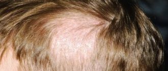
- bald spots on the head in the hairline area (clipping form);
- fever (rare);
- malaise, lethargy.
Symptoms of the disease
When infected with pityriasis versicolor, the pathogens multiply in the superficial layers of the skin, disrupting the normal functioning of cells. First of all, melanocytes, which are responsible for the production of the pigment melanin, are affected, thanks to which the skin has one color or another. This means that under the influence of pathogenic microorganisms, the affected areas acquire an uncharacteristic, painful shade. Hence the second name for pityriasis versicolor - versicolor.
The main symptoms of pityriasis versicolor include:
- Spots of yellow, coffee, scarlet or dark brown shades, located mainly on the back, head and neck, shoulders and stomach. In children, they also appear on the limbs, armpits and scalp. The affected areas can be of different sizes, but usually their size does not exceed 1 cm. Subsequently, the spots merge with each other, forming significant areas of the affected skin.
- No signs of inflammation in the form of redness, swelling, hyperemia and pain to the touch.
- Severe peeling of the skin in infected areas, aggravated by touch.
- No tanning on the affected areas.
Pityriasis versicolor is not characterized by itching and discomfort, so patients often mistake the manifestations of the disease for minor cosmetic defects, postponing a visit to the doctor and full treatment.
Such spots begin to itch and scratch only if a secondary infection occurs.
Reasons for development
Versicolor versicolor is a type of fungal skin infection that affects the stratum corneum of the epidermis and hair follicles. Its causative agents are two types of fungi, and infection is possible only through prolonged and close contact with the patient. And in this case, provoking factors play a big role. These include:
- weakened immunity;
- hyperhidrosis;
- disruption of the sebaceous glands;
- diseases of the endocrine system (obesity, diabetes, Itsenko-Cushing syndrome, etc.);
- hormonal imbalance due to pregnancy, menopause or taking hormone-containing medications;
- vegetative-vascular dystonia;
- abuse of antibacterial personal hygiene products;
- excessive exposure to ultraviolet rays (intense tanning, frequent visits to the solarium) and regular overheating of the body.
It is noteworthy that patients with pityriasis versicolor over 60 years of age are extremely rare. This is due to natural age-related changes in the skin, which make it less susceptible to pathogens.
In children under 10 years of age, the main causes of pityriasis versicolor infection are neglect of personal hygiene rules or improper skin care. At this age, with the protective functions of the skin intact, the body independently copes with pathogenic microorganisms attacking it, so the development of the disease does not occur. But closer to adolescence, when hormonal changes begin, the body’s susceptibility to bacteria, viruses and fungi increases, so children over 10 years old become infected with pityriasis versicolor just like adults.
HERPES SIMPLE (HERPESIS SIMPLE)
This paper outlines the present-day concepts of the pathogenesis of herpes simplex, describes its main clinical manifestations, and considers its therapy. V.N. Grebenyuk, doctor med. Sciences, prof., head. Department of Pediatric Dermatology of the Central Research Institute of Dermatovenerology of the Ministry of Health of the Russian Federation. VN Grebenyuk, professor, MD, Head, Department of Pediatric Dermatology, Central Research of Dermatovenereologic Institute, Ministry of Health of the Russian Federation. P
Herpes growth is a serious medical and social problem. This is one of the most common human viral infections, often characterized by a persistent chronic course, affecting various organs, systems and tissues. According to WHO, about 70% of the population of our planet is infected with the herpes simplex virus (HSV) and approximately 10 - 20% of those infected have some clinical manifestations of herpes infection. HSV is a predominantly dermatoneurotropic DNA-containing virus; it also has tropism for other tissues, its size is 150 - 300 nm. The virion, in addition to DNA, consists of an icosahedral capsid and an outer shell containing lipids. It reproduces intracellularly (in the nucleus and cytoplasm) with a 14-hour reproduction cycle. During an acute infectious process, daughter virions are released from decaying cells. HSV infection can cause spontaneous abortions, fetal death and congenital deformities. The herpes virus is associated with the possibility of developing cervical cancer and some cardiovascular diseases. There are two antigenic types HSV-I and HSV-II, which cause lesions of the skin and mucous membranes of various localizations, which is determined by the place of introduction of the virus, usually through contact (coitus, kissing, through household objects). The source of infection can be not only patients with herpes, but also virus carriers who do not have symptoms of herpes.
| Rice. 1. Herpetic lesions of the face. a - forehead, eyelids, bridge of the nose; b - cheeks; c - lips and chin. |
3-4 weeks after infection, antibodies to HSV are formed in the body, the level of which remains relatively constant throughout a person’s life, regardless of the form of infection - manifest or latent. In the vast majority of people, the infection is asymptomatic or subclinical, and only in some infected people does it manifest clinically. Having penetrated the body, the herpes virus reaches a certain regional sensory ganglion (spinal or cranial) through lymphogenous, hematogenous or neurogenic routes, where it constantly persists. The latent state of the virus is based on the biological balance between micro- and macroorganisms. Under the influence of various provoking factors (psycho-emotional arousal, intoxication, overheating, etc.), a relapse of the disease occurs due to the reactivation of latent HSV, which leads to the formation of a recurrent disease. The range of clinical manifestations of the disease - from virus carriage to generalized forms - is determined both by the biological properties of the pathogen and the reactivity of the host. In most people, immune mechanisms, mainly cellular, maintain HSV latency. But in some infected people, antiviral resistance turns out to be untenable and relapses occur. There are two hypotheses that allow for the development of relapses based on both the static and dynamic state of the virus. According to the first hypothesis, the virus is located in the cells of the paravertebral sensory ganglion in an integrated or free non-productive state. Under the influence of the “trigger factor,” the virus, when activated, moves from the ganglion along the axon of the peripheral nerve to the epithelial cells, where it replicates. Cell susceptibility and weakened immune control are thought to contribute to this. According to the dynamic state hypothesis, replication and release of small amounts of virus from the ganglion occur continuously. Reaching the skin nerve, HSV causes microfoci of infection, which are restrained by defense mechanisms, which prevents relapses or weakens their manifestations. The development of relapses is also influenced by the state of local immunity. Its inhibition creates conditions for the replication of the virus that has reached the skin. The immune system plays an important role in containing the spread of herpes infection in the body. Immune protection is determined by the interaction and complex participation of specific and nonspecific factors. The main place in this system belongs to T-cell mechanisms of immunity. Mononuclear phagocytes and neutrophils play a significant role in maintaining local immunity and preventing the dissemination of infection. The protective functions of the body and the preservation of its homeostasis are also greatly influenced by the ability of cells to produce interferon.
Rice. 2. Herpetic felon.
| Rice. 3. Manifestations of herpes. a - on the palmar surface of the hand; b - on the thigh; c - on the buttocks |
Diseases caused by HSV are distinguished by a wide clinical variety of localization, severity, and characteristics of clinical manifestations. Primary herpes usually occurs after the first contact with HSV. More often it is observed in childhood against the background of a reduced immune status, in particular in the absence or low content of specific humoral antibodies. It is distinguished by the high intensity of clinical symptoms. The incubation period lasts several days. Primary herpes in newborns due to hematogenous dissemination becomes systemic, affecting the central nervous system and internal organs. The disease is characterized by herpetic lesions of the oral cavity, eyes, liver, bronchi, lungs, and brain. Usually the disease occurs acutely in the first days after birth and is manifested by anorexia, dyspeptic disorders, convulsions, septic condition, body temperature (39 - 40 ° C), disseminated herpetic rash on the skin and mucous membranes; Deaths are common in the first 2 weeks of illness. Children who have had generalized herpes experience neuropsychic complications. Kaposi's eczema herpetiformis is another severe type of herpes. Occurs mainly in children. It usually occurs in patients with atopic dermatitis, eczema, and other dermatoses in which there are skin lesions. The source of the disease can be patients with herpes in the acute stage. In adults, the disease may be associated with a recurrence of herpes labialis or another clinical form. Kaposi's eczema herpetiformis is characterized by a sudden onset (chills, malaise, body temperature up to 39 - 40 ° C for 1 - 1.5 weeks), a profuse vesicular rash on large areas of the skin, and painful regional lymphadenitis. The rashes appear in paroxysms over 2-3 weeks at intervals of several days. Often, along with skin lesions, the mucous membranes of the oral cavity, pharynx, trachea, and eyes are involved in the infectious process. Grouped and disseminated vesicles soon turn into pustules. In the center of the rash elements there are often umbilical recesses. After the crusts are rejected, secondary erythema remains on the vesiculopustules. Subjectively, the rash is accompanied by itching, burning, and soreness of the skin. Regional lymphadenitis is not uncommon. Patients are subject to hospitalization in an infectious diseases hospital or clinical hospital wards. In severe forms, the pathological process may involve the nervous system, eyes and internal organs. Relapses of Kaposi's eczema herpetiformis are rare, characterized by shorter duration and weakened clinical manifestations.
| Rice. 4. Genital herpes. a — bubble manifestations; b — erosive and ulcerative manifestations. |
The most common clinical form of primary infection is acute herpetic stomatitis.
It is more often observed in children in the first years of life; it is rare in adults. In weakened children, dissemination of the virus can lead to visceral pathology (in particular, hepatitis) and death. Acute herpetic stomatitis, occurring after about a week's incubation period, is characterized by a violent clinical picture. Chills, high body temperature (up to 39° C), painful vesicular-erosive rashes in the oral cavity, headache, general malaise, drowsiness - this is a list of the main symptoms of this disease. The rashes are most often located on the mucous membrane of the cheeks, gums, palate, lips, tongue, less often - on the soft and hard palate, palatine arches and tonsils, and spread to the skin around the mouth. The rash initially looks like grouped vesicles against a background of erythematous-edematous islands of the mucous membrane. The transparent contents of the elements become cloudy after 1 - 2 days, the covers of the vesicles are destroyed, and erosions form. In this case, regional lymph nodes are almost always enlarged and painful. Regression of the process usually occurs after 2 - 3 weeks. Recurrences of herpetic stomatitis, as a rule, are milder and resolve earlier. Herpes simplex is more common as a recurrent form. Clinical manifestations compared to primary herpes are less pronounced and not as long lasting. Most often, rashes are located on the face (lips, cheeks, nose), conjunctiva and cornea of the eyes, on the genitals and buttocks. The disease can last for many years and recur with varying frequencies - from several times a year to several times a month. In rare cases, the process becomes permanent when new rashes appear against the background of previous lesions that have not yet resolved. Frequent relapses of genital herpes are especially painful. The localization of herpetic lesions is determined by the site of virus introduction. The appearance of the rash is preceded by prodromal symptoms (burning, itching, tingling and other sensations). Grouped vesicles with a diameter of about 2 mm occur against a background of erythema. The transparent contents soon become cloudy and shrink into lumpy-yellowish crusts. When the vesicles rupture, scalloped erosions form. Their bottom is soft, reddish, the surface is smooth and moist. Regional, slightly painful lymphadenitis with a pasty consistency often occurs. The rash resolves within 1 to 2 weeks, leaving reddish-brown spots. When a microbial infection is added, the duration of relapses increases. Atypical forms of herpes simplex are known: abortive, zosteriform, disseminated, hemorrhagic-necrotic, migratory, elephantiasis-like, ulcerative, rupioid. The abortive form occurs in areas of the skin with a thickened stratum corneum and manifests itself as barely noticeable papules. Abortive manifestations of the disease also include erythematous and pruriginous-neurotic forms, characterized by local subjective disorders without typical rashes. The edematous form is usually located in areas of the skin with loose subcutaneous tissue (eyelids, lips) and is characterized by pronounced tissue swelling. Zosteriform herpes simplex is localized along the course of a nerve on the limbs, trunk, face and is accompanied by neuralgia, headache and general weakness. In the disseminated form of the disease, the rash simultaneously appears on areas of the skin that are distant from each other. The migratory form of recurrent herpes is characterized by a change in the localization of lesions. In hemorrhagic and hemorrhagic-necrotic forms, an admixture of blood is detected in the contents of the vesicles and necrosis develops. The elephantiasis-like form of the disease is characterized by severe swelling followed by the development of persistent elephantiasis in the affected area. Chronic cutaneous herpes simplex is an extremely rare clinical form. It is observed in patients with immunodeficiency and is characterized by permanent active manifestations of infection. Persistent ulcerative lesions up to 2 cm in diameter appear. The ulcerative form of herpes simplex is characterized by the development of ulcerative lesions, which is associated with a weakening of the patient’s immunobiological defense mechanisms and the increased virulence of the virus strain. This clinical type of herpes is characterized by the formation of ulcers at the site of weeping vesicles and fused erosions. The bottom of the ulcers is soft, pink-red in color, sometimes with a grayish-yellowish coating. In the first days of the disease, local pain and burning are expressed. Sometimes the rash is accompanied by inguinal lymphadenitis. The rupoid form of herpes simplex is usually localized on the face. It is caused by pyogenic infection with the development of cracks and layered crusts. Relapses occur several times a year. The rash is often accompanied by tenderness and enlargement of regional lymph nodes. With herpes of the hands, the process is often located on the distal parts of the hands. Limited lesions are represented by single dense blisters, accompanied by severe pain. The most common type of herpes simplex is facial herpes. In most people, these are sporadic focal vesicular eruptions, often resolving within 1 week. In severe cases, the process involves large surfaces of the face - nose, cheeks, forehead, skin and red border of the lips. Genital herpes occupies a significant place in the structure of herpetic diseases. Etiologically, its occurrence is equally often associated with types of HSV-I and/or HSV-II. Infection with one type of virus does not prevent the occurrence of HSV infection of another type, which leads to the formation of intermediate (“double”) antibodies. Mixed infection with HSV-I and HSV-II is quite common. The frequent isolation of HSV-I, which was previously considered the causative agent of non-genital forms of herpes, in genital lesions is due to the prevalence of orogenital contacts. Genital herpes is distinguished by the variability of its clinical picture and its tendency to have a chronic, relapsing course. In men, limited herpetic eruptions are often localized on the inner layer of the foreskin, in the head groove, and less often on the head and shaft of the penis. In women, the labia minora, clitoris, cervix, perineum and thighs are most often affected. Rashes (vesicles, erosions, ulcers, cracks) against a background of erythema and swelling are usually painful and are also accompanied by itching, a feeling of tension and heaviness in the perineum. About a third of patients have inguinal lymphadenitis. When the urethral mucosa is involved in the pathological process, serous discharge from the urethra and pain when urinating appear. The source of infection in the case of genital herpes is usually a patient in the acute stage of the disease; it can also be a virus carrier, given the possibility of asymptomatic persistence of HSV in the genitourinary tract in men and in the cervical canal. The incubation period for primary genital herpes lasts from one to several days. Clinically, primary genital herpes has a more severe and prolonged course. The localization of rashes on the genitals and adjacent areas is determined by the gates of the viral infection. A recurrent course of genital herpes is observed in the majority of infected people. Provoking factors are a variety of influences - psycho-emotional experiences, hypothermia, menstruation, weather and climate fluctuations, and other factors that disrupt the state of biological balance of the body, contributing to a decrease in the immune response and activation of HSV. The clinical picture, the amount of virus secreted by the patient and the associated infectivity are more pronounced with primary herpes than with a recurrent disease. Possible complications of herpes simplex: the addition of a secondary bacterial infection, reinfection with the released virus of other epithelial integuments, neurological manifestations (aseptic meningitis, transverse myelitis), encephalitis, disseminated infection of internal organs, psychosocial consequences (psychological instability). The risk of developing cervical cancer is 2 times higher in women who are seropositive for human papillomavirus types 16/18 and infected with HSV-II.
Diagnostics
The diagnosis of herpes simplex, especially its genital form, in most cases is based on the clinical picture. Difficulties arise with atypical manifestations of herpes. In this case, it is important to carefully collect anamnesis, paying attention to relapses accompanied by itching, burning, and ineffectiveness of antibiotic therapy. In addition, the patient may have a tendency to colds, general weakness, malaise, low-grade fever, and depression. Recurrent herpes is characterized by a wave-like course of the disease - an alternation of relapses and remissions. In women, relapses of herpes may be associated with certain phases of the menstrual cycle. The occurrence of erosions and ulcers on the genitals simulates syphilitic lesions. This similarity is most pronounced when a secondary microbial infection is attached, as well as during irrational therapy. The diagnosis of genital herpes is complicated by the fact that HSV is often associated with some resident autoflora microorganisms: chlamydia, streptococci and staphylococci, gardnerella and others, which can determine the occurrence of mixed infections. In addition, because herpes can be transmitted sexually, the patient must be tested to rule out other sexually transmitted diseases, including syphilis and AIDS. In complex cases, when clinical data is insufficient, laboratory diagnosis is possible. There are a number of specific laboratory tests to recognize HSV infection: isolation of HSV in cell culture, including HSV-I and HSV-II typing, tests to determine HSV antigen or DNA using polymerase chain reaction; serological tests - complement fixation test, ELISA, indirect immunofluorescence reaction, reverse passive hemagglutination reaction, protein-specific immune tests (immunoblotting), cytological examination (detection of multinucleated giant cells in scrapings from the lesion).
Treatment
Treatment of recurrent herpes remains a difficult task, which is not always solved effectively. It is possible to achieve some success if complex etiological and pathogenetic treatment is carried out at different stages of the disease, aimed, on the one hand, at suppressing the infectious agent, and on the other, at increasing the body’s immune reactivity. When choosing treatment, the stage of the disease should be taken into account. For relapses, interferon, antiviral chemotherapy, measles immunoglobulin, human normal immunoglobulin, levamisole, ascorbic acid, deoxyribonuclease, applications of 0.05% zinc sulfite solution are indicated; in the inter-relapse period - herpetic and polio vaccines, pyrogenal. The etiological focus is on antiviral chemotherapy drugs, which are more effective when used in the first hours and days of the appearance of rashes. Among them is the domestic drug Bonafton, which is used orally at 50–150 mg/day for 5–7 days for relapses. Simultaneously with the tablet form, 0.5% bonaftone ointment can be prescribed. It is applied to the lesions in an open manner when signs of relapse appear and is easily rubbed into the skin 2 - 3 times a day for 5 - 7 days. Side effects observed in some patients include malaise, loose stools, and dermatitis. Acyclovar (Zovirax) is effective, characterized by low toxicity and selectivity against HSV. The drug is used intravenously, orally and topically. It gives a pronounced therapeutic effect for Kaposi's eczema herpetiformis. Acyclovir is administered intravenously at the rate of 20 mg per 1 kg of body weight per day. However, the drug does not prevent herpes from recurring, infecting newborns, or infecting other people. Treatment of patients with recurrent herpes with acyclovir 0.1 - 0.2 g 5 times a day for 5 days during relapses shortens the time for resolution of rashes, reduces the severity of subjective sensations, smoothes out clinical manifestations and reduces the degree of virus shedding. Prophylactic administration of the drug 0.1 - 0.2 g 4 times a day for 6 - 12 weeks reduces the duration of relapses and weakens clinical manifestations. Other chemotherapy drugs: famciclovir, alpizarin (2 and 5% liniment), Viru Merz Serol, 1% oxolinic ointment, hevisos, ribavirin (virazol). A certain therapeutic effect is provided by immunocorrective drugs (myelopid, poludanum, arbidol), used both as monotherapy and in complex treatment. Myelopid (0.003 g in 2 ml of saline) is administered intramuscularly once every 3 days (5 injections per course). Treatment is carried out in two courses with an interval of 7 - 10 days. Poludan is administered subcutaneously into the forearm every other day, 100 mcg, for a course of 1000 mcg. Arbidol is prescribed 0.2 (2 tablets) 3 times a day - 5 days with a 2-day break, and then 0.1 g (1 tablet) 1 time per week for 3 weeks. Sodium nucleinate is also used orally at 0.5 - 1 g / day in 2 - 3 doses daily for 2 - 4 weeks. Taktivin is used to stop relapses and for prophylactic purposes. The drug is administered subcutaneously at a dose of 100 mcg every other day, 8 - 10 injections. During the inter-relapse period, 50 mcg is prescribed every other day, a course of 5 injections is repeated every 3-6 months. A course (4 - 5 injections) of treatment with timoptin is also carried out, which is administered subcutaneously at 100 mcg every 3 - 4 days. The courses are repeated after six months.
External treatment
Antiviral ointments, creams, lipsticks accelerate the epithelization of erosions, reduce or reduce subjective sensations in the affected areas. Local use of one or another antiviral drug in the treatment of herpetic lesions for 5 - 7 days shortens the time of regression; use 2 - 3 times a week during the inter-relapse period allows to prolong remission. Interferon has an inhibitory effect on HSV, which is applied to the skin and easily rubbed in for 4 to 7 days. During treatment, it is advisable to alternate antiviral drugs during relapses. Human interferons are effective in the treatment of recurrent herpes in the prodromal period and when the first signs of relapse appear. The ointment is applied to the lesions 2-4 times a day and rubbed in lightly; treatment is continued for a week. The use of interferon ointment during the inter-relapse period prolongs remissions and interrupts the development of relapses. In order to prevent relapses in frequently recurrent forms of herpes, patients for whom treatment is ineffective are prescribed a herpetic vaccine. Contraindications to its administration are lesions of parenchymal organs, diabetes mellitus, stage II and III hypertension, decompensated heart failure, acute infections and allergic diseases. The drug is administered intradermally during the period between relapses, 0.2 - 0.3 ml into the area of the flexor surface of one of the forearms. The first 5 injections are given after 3 - 4 days, the next 5 doses are administered after a 2-week break (once every 5 - 7 days). These 10 injections constitute the main course of treatment, 3–6 months after the end of which 1–2 cycles of revaccination are carried out, each of 5 injections with an interval between injections of 7–14 days and between cycles of 6–8 months. Over the next 2 years, an additional revaccination cycle of 5 injections is carried out every 8 - 12 months. At the injection site, after 18-24 hours, a local reaction develops, manifested by the development of erythema with a diameter of 2-5 cm with a papule in the center and accompanied by a burning sensation. During vaccination, a focal reaction such as abortive relapses may be observed. In this case, a break is taken in the treatment for 2 - 3 days, then it is continued. Specific vaccine therapy leads to an increase in the duration of remissions, a reduction in relapse periods, and the disappearance of subjective sensations. For the purpose of secondary prevention of relapse of herpes, the factors that provoke the disease are controlled. Great importance is attached to the sanitation of the body and health-improving measures in the process of medical examination.
Literature:
1. Barinsky I.F., Shubladze A.K., Kasparov A.A., Grebenyuk V.N.M.: Medicine. 1986, 269 p. 2. Masyukova S. A., Rezaikina A. V., Grebenyuk V. N., Fedorov S. M., Mkhitaryan A. G., Kolieva M. Kh. Immunotherapy of recurrent herpes simplex. Sexually transmitted diseases. Information analytical newsletter. Sanam Association 1995, 3, 27-30. 3. Minde CA. Genital Herpes. A guide to pharmacological therapy. Drugs 1994;47(2):297-304. 4. Whatley JD, Thin RN. Episodic acyclovir therapy to abort recurrent attacks of genital herpes simplex infection. J Antimicrobial Chemotherapy 1991;27:677-81.
Treatment of pityriasis versicolor
The diagnosis of pityriasis versicolor is made to the patient after examination by a dermatologist and dermatoscopy. Additionally, an iodine test and laboratory testing of scrapings can be used.
Treatment of pityriasis versicolor is carried out on an outpatient basis until the symptoms of the disease completely disappear. If measures were taken on time, the patient is prescribed local therapy using antifungal ointments and special agents for exfoliating dead cells.
Additionally, immunomodulators, vitamin complexes, antifungal shampoos and antihistamines are used if the patient is bothered by itching. In the most advanced cases, antimycotic agents are prescribed for oral administration. In addition, in order to avoid relapse of the disease and infection of others, the patient’s clothing and bedding are treated with disinfectant compounds.
Disease prevention also plays an important role. The following will help prevent the development of the disease:
- timely solution to the problem of hyperhidrosis (use of medicinal deodorants, creams, powders, compliance with personal hygiene rules, frequent changes of underwear, etc.);
- use of high-quality soaps and skin care products;
- regular water procedures;
- wearing clothes made from natural, hypoallergenic materials;
- avoiding stress;
- balanced diet rich in vitamins and microelements.
Experts also recommend avoiding overheating of the body and promptly seeking advice from cosmetologists and dermatologists in order to identify the problem and begin proper treatment.
What can be confused with pityriasis alba?
In some cases, it is necessary to differentiate lichen alba with the following pathologies:
- vitiligo;
- contact and atopic dermatitis;
- post-inflammatory hypopigmentation of any origin (including after the use of drugs);
- fungal infections;
- shingles;
- depigmentation of nevus (anemic nevus);
- psoriasis;
- seborrhea;
- pityriasis rosea;
- discoid and nummular eczema [1,3].
What is erythrasma
Erythrasma is a chronic bacterial disease affecting the epidermis layer in the deep folds of the skin. It is characterized by a long course - in some cases, symptoms develop for at least 10 years, without causing significant discomfort to the patient. The clinical picture of erythrasma is similar to a fungal infection of the skin, but modern dermatology classifies it as a group of pseudomycoses.
The following main stages are distinguished in the development of the disease:
- Progression. The first characteristic spots appear on the skin, their size slowly increases, and additional symptoms develop. In some cases, secondary infections occur. The spots gradually merge with each other, forming large areas of damage.
- Stabilization. New spots do not appear, and existing ones stop growing. Peeling of the skin begins. This stage is usually associated with a change in external conditions, for example, cold weather, during which the intensity of sweating decreases and the skin condition stabilizes.
- Exacerbation or relapse. Usually associated with the beginning of the warm season. But in the case of prolonged erythrasma, the disease constantly develops in waves, and after a slight decline its symptoms again actively appear.
- Remission. Occurs with a favorable microclimate, compliance with preventive measures and proper skin care. The color of the affected areas gradually returns to normal, itching and flaking disappear, and the skin is restored.
Without timely, well-chosen treatment, erythrasma can lead to the development of serious complications.
For example, it can provoke eczema and secondary infection in patients with diabetes or obesity. Also, the course of the disease is aggravated by increased humidity and contamination of the affected areas. As a result, its typical symptoms are complicated by burning, itching and pain.
Signs of the disease
Externally, erythrasma manifests itself in the form of light brown, brick-red, brown or yellow-brown spots on the skin, most often round in shape and without signs of inflammation. The diameter of the lesions can reach several centimeters, and they tend to merge, forming large affected areas. First of all, erythrasma spots appear in the folds of the skin, where there is a favorable environment for the proliferation of bacteria.
In addition to spots, erythrasma is characterized by:
- Peeling of the skin on the affected areas, aggravated by touch. Usually this is where the development of the disease begins.
- Mild, irregular itching. It intensifies and begins to cause significant discomfort only if a secondary infection is added to the primary disease.
- Absence of fever, wounds, ulcers and ulcers with copious discharge. This distinguishes erythrasma from most bacterial skin pathologies.
- Getting wet. An optional symptom, the manifestation of which depends on the amount of sweating and the quality of skin care.
It is noteworthy that in children, symptoms of erythrasma appear extremely rarely. The risk group includes adults, primarily men, who are predominantly overweight and prone to excessive sweating. In this case, in men, the skin in the groin, navel and inner thighs is usually affected, and in women, the entire abdomen, armpits and areas under the breasts are affected.
Causes of pathology
Corynebacteria, which are the causative agents of the disease, are normally always present on human skin. Moreover, they provoke the development of pathology only under certain, favorable conditions. Corynebacteria do not penetrate deeper than the epidermis, and also do not affect nails and hair. Since the appearance of erythrasma is directly related to increased sweating, the disease most often manifests itself in the hot summer season.
Among the main reasons for the development of erythrasma are:
- hyperhidrosis;
- deviation of the normal pH of the skin to the alkaline side;
- diaper rash, constant friction and mechanical damage to the skin;
- dermatitis and other skin diseases;
- neglect of personal hygiene rules;
- wearing synthetic, overly warm clothing;
- the use of low-quality care products or the abuse of soap with an antibacterial effect, which destroys the natural protection of the skin.
Erythrasma is transmitted by contact, most often after the use of clothing, bedding and personal hygiene products of the patient. You can also become infected during sexual intercourse, when visiting a pool or bathhouse, and when walking barefoot on the ground or beach. At the same time, it is not always possible to accurately determine the source of infection, because the carrier may not have obvious external manifestations of the disease in the form of characteristic spots and peeling.
Treatment of erythrasma
To diagnose a patient with erythrasma, a dermatologist first uses a visual examination. This is especially true for rashes in the groin area, which have characteristic distinctive features in the form of pronounced protrusions and bubbles along the edges. Also, the affected areas of the skin are illuminated with a Wood's lamp and a microscopic examination of the scraping is performed to exclude other diagnoses: pityriasis versicolor or pityriasis rosea, candidiasis, dermatitis or eczema.
Treatment of erythrasma is primarily based on the use of antibacterial ointments that are used to treat the affected areas of the skin.
Under their influence, corynebacteria die, and the spots gradually lighten, decrease in diameter and disappear. On average, such therapy takes at least 7-10 days. Used in parallel:
- Antiseptics. Treatment with them is carried out before applying antibacterial ointment, as well as after it, to maintain dryness of the affected areas and prevent re-infection.
- Antifungal drugs. They are prescribed together with antibacterial drugs, since corynebacteria are similar in structure to fungal micelles.
- Exfoliating ointments. They help cleanse the skin of a layer of dead cells, activating its regeneration.
- Ultraviolet irradiation. Promotes skin disinfection and restoration. Patients benefit from both natural sunbathing and physiotherapeutic UV irradiation.
If the disease has not reached an advanced stage, the use of external medications is sufficient to solve the problem. But in some cases, with multiple skin lesions, to obtain the desired result, patients are prescribed systemic antibacterial therapy.
Diagnosis of lichen alba
Patients turn to doctors on their own, as in most cases they are concerned about the appearance of unusual rashes that cause them anxiety. Usually they turn directly to dermatologists, less often to pediatricians or therapists. Making a diagnosis, as a rule, does not cause difficulties for a specialist, since the symptoms of pityriasis alba are very characteristic. Already at the first appointment, without resorting to additional examination methods, the dermatologist determines this type of lichen.
How can the diagnosis be confirmed? For this, additional types of diagnostics are used:
- examination of the spot under a Wood's lamp to exclude vitiligo: lichen alba does not fluoresce, the color of the spot does not change, hypopigmentation does not increase;
- Dermatoscopy for this type of lichen reveals an area with unclear boundaries and slight peeling, the hair color in the area of the spot is the same as on the rest of the body;
- reaction with KOH solution to detect fungal infections is negative;
- microscopy and culture for mycoses are negative;
- A biopsy is not usually performed, but if done, a decrease in the number of melanocytes and the absence of traces of infectious agents can be detected [2,5].